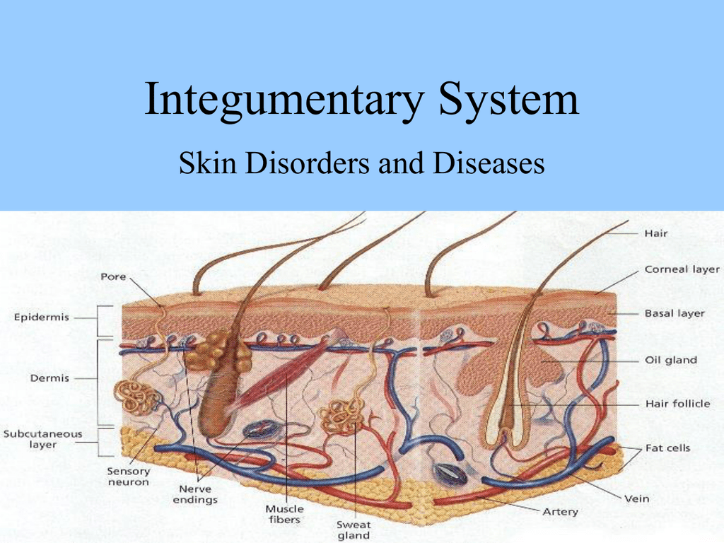
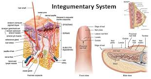
ENGLISH-integumentary -PART-1(UPLOADED)
INTEGUMENTARY SYSTEM DISEASES AND DISORDERS (SKIN)(Integumentary System Diseases and Disorders):
Nursing Management of patient with
diseases and disorders of integumentary
system
a) Nursing Assessment
History
Physical assessment
b) Etiology
c) Pathophysiology
d) Clinical manifestations
e) Nursing management of disorders of skin
and its appendages
- Lesions and abrasions
- Infection and infestations Dermititis
- Dermatoses; infectious and non
infectious - Inflammatory dermatoses
- Acne Vulgaris
- Allergies and Eczema
- Psoriasis
- Malignant Melanoma
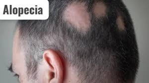
- Alopecia
- Infestations
- Bacterial infections
- Pyoderma
- Impetigo
- Folliculitis
- Furuncles
- Carbuncles
- Viral infections
- Herpes zoster
- Herpes simplex
- Fungal infection
- Athlete’s foot (Tanta Pedi’s)
- Parasitic infestation
- Pediculosis
- Scabies
- Pemphigus
- Stevens-Johnson syndrome
- Skin cancer
- Special dermatological therapies
f) Burn and its management - Burns Plastic Surgery
- Incidence, causes of burns
- Types & classification of burns
- Pathophysiology
- Calculation of the percentage
- Local & systematic effects of burns
- Immediate care
- First aid care
- Medical management, barrier nursing
care of the burns - Complications, Health education
g) Plastic Surgery - Define plastic & reconstructive surgery
- Types
- Define skin graft flaps
- Possible complication
- Preparation of patient for constructive
surgery - Post operative care
- Health Education
h) Alternate therapies
i) Drugs used in treatment of integumentary
disorders
Important Terminology for disorders of integumentary
system:
Alopecia :- Loss of hair
Acantholysis :-
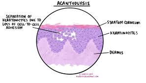
- Epidermal cells separating from each other (which occurs due to damage or abnormality of intracellular substances)
Carbuncle (carbuncle) :-
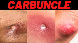
- A carbuncle is a bacterial skin infection that affects a group of hair follicles.
Cellulitis (Cellulitis) :-
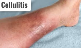
- Cellulitis is a bacterial skin infection. In which redness, swelling and pain are seen in the affected skin area.
Comdones (comdones) :-
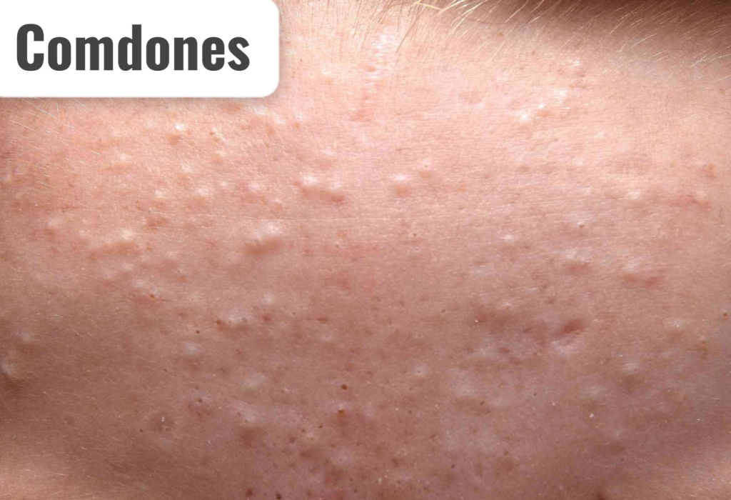
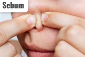
- A comedon is a skin-colored papule-like formation that is found in acne vulgaris. Which is seen due to sebum blockage in the hair follicle.
Cryosugery (cryosurgery) :-
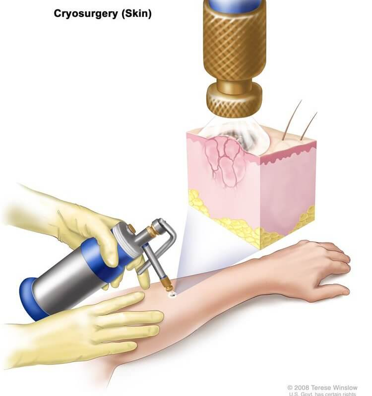
- Cryosurgery is a process in which liquid nitrogen is used to produce extremely cold temperatures that destroy cancerous cells and abnormal tissue.
Cynosis :-
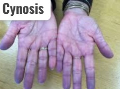
- Bluish discoloration of skin and mucous membranes.
Cytotoxic:-
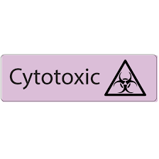
- A cytotoxic is a substance or procedure that causes cell damage and cell death.
Dermatitis :-
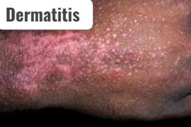
- Inflammation of skin
Dermatosis :-
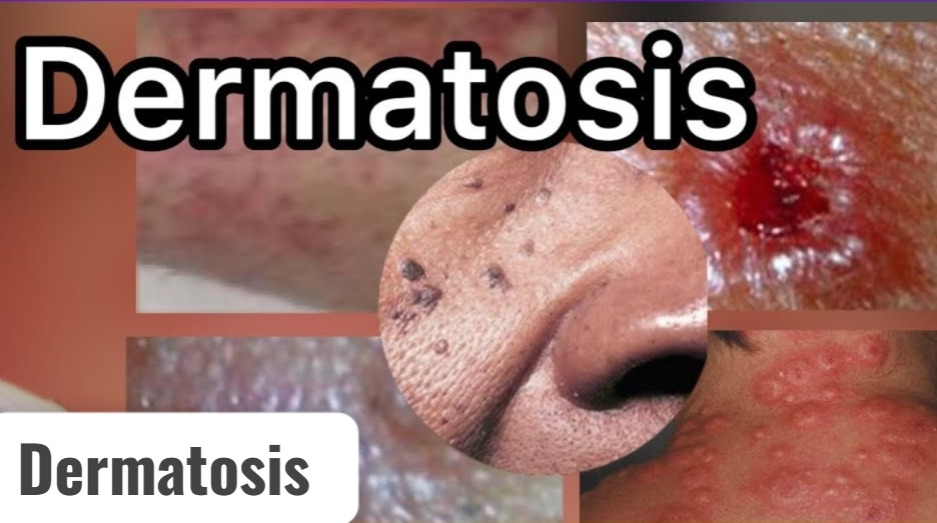
- Abnormal skin lesion
Debriment :-
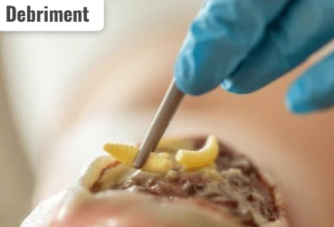
- Necrotic and infected tissue Removal Procedure.
Dermatophytoses :-
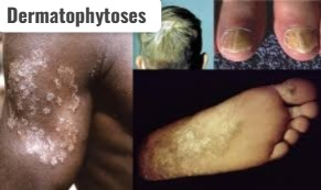
- Fungal infection of skin
Erythema :-
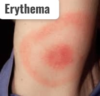
- Red color of the skin due to congestion of capillaries.
Erysipelas :-
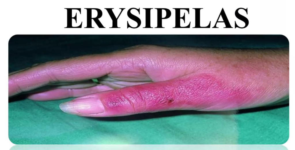
- Erysipelas is a bacterial skin infection. In which the superficial dermis layer of the skin is affected.
Folliculitis (folliculitis) :-
- Bacterial infection of the hair follicle is called folliculitis. (Infection of hair follicle)
Furuncle :-
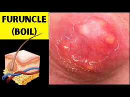
- A furuncle is a bacterial hair follicle infection. In which painful, pus-filled bumps are seen under the skin.
Hydrophilic (hydrophilic) :-

- Hydrophilic is a type of material that absorbs moisture.
Hydrophobic (hydrophobic) :-

- Hydrophobic is a type of material that repels moisture.
Hygroscopic (Hygroscopic) :-

- Hygroscopic is a type of material that absorbs moisture from the air.
Hirsutism :-

- Hirsutism is excessive hair growth in women.
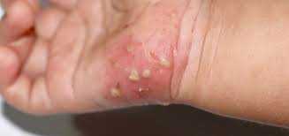
Impetigo (Impetigo) :-
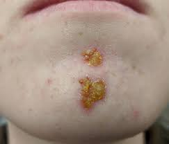
- Impetigo is a common bacterial infection that causes sores and blisters on the skin.
Keratin (Keratin) :-
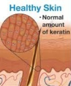
- Keratin is a fibrous protein that forms the outer layer of the skin.
Keloids :-
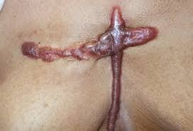
- Kiloids irregular, thick Scars. Which are seen due to abnormal wound healing process.
Melanin :-
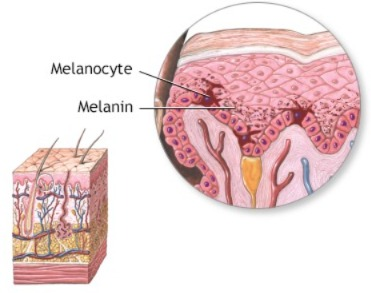
- Melanin is a substance. which is responsible for skin color.
Melanocyte :-
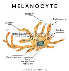
- A melanocyte is a skin cell that produces melanin.
Patechia (Petechia) :-
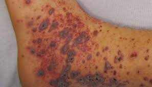
- Petechiae are red spots seen on the skin. Which is seen due to blood leakage in the skin.
Plasmapharesis (Plasmapharesis) :-
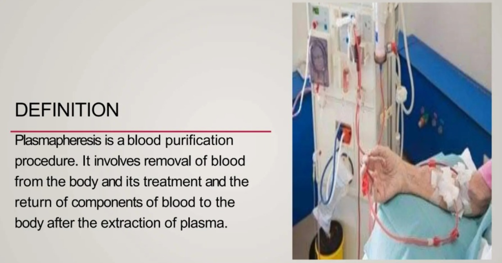
- Plasmapheresis is a process in which bad plasma is removed from the blood through centrifugation and reinfusion and new plasma is added in its place.
Psoriasis (Psoriasis) :-
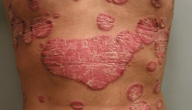
- Psoriasis is an autoimmune disorder in which epidermal cells in the skin are produced rapidly.
Pruritus (pruritor) :-

- Pruwriters is the term used for etching, which causes scratches.
Paronychia :-
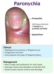
- Infection of the soft tissue around the fingernail and toenail is known as paronychia.
Scabies :-
- Scabies is a parasitic infestation. Which is caused by the Sarcoptes scabiei mite.
Sebum :-
- The fatty secretion produced by the sebaceous glands is known as sebum.
Telangiectasia (Telangiectasia) :-
- Superficial and small veins in the skin become dilated.
Tinea:-
- Fungal Infection of Skin and Scalp.
Ulticaria:-
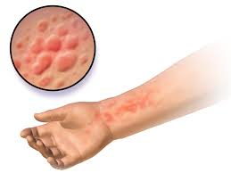
- Uticaria is a type of itchy rash. Which is seen due to a reaction to food, medicine or any other substance.
vitilogo (Vitilogo):-
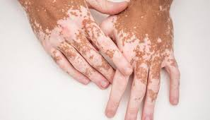
- In vitiligo, the cells that produce pigmentation die or stop working, causing the skin to lose its color and appear excessively white.
Warts :-
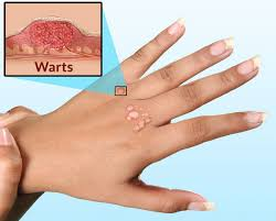
- Warts are a condition caused by a virus in the human papillomavirus. In which a small bump is seen on the skin.
Wood light (Wood light):-
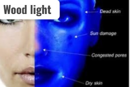
- Wood light is a blue type of light used for skin assessment. So that abnormal skin conditions can be identified.
Xerosis (Xerosis):-
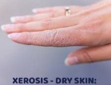
- Dry skin
a) Nursing Assessment :-
History :-
(A) Present health history :
- Ask the patient about their current complaints. Ask the patient if there is any itching, dryness, redness, lesion lump, swelling present in any area. Ask about any symptoms found on the skin, hair, nails, or scalp.
- Ask about when the symptoms started and ask about their duration, intensity, and location.
Past health history :
- Collect information about previous sun and radiation exposure.
(B) Past surgical history :
- Collect information about any cosmetic surgery or any other type of surgery performed in the past years To do.
(C) Personal history :–
- About whether the patient has any skin allergies or not Ask.
- Ask about any allergic reactions to any food, medicine or chemical.
- Know about the cosmetic items, soaps, shampoos and personal hygiene products used by the patient.
- Know about the patient’s elimination pattern, sleep-rest pattern, sexuality pattern, exercise and activity pattern.
- In addition, know about the type of material the patient uses in the clothes.
(D) Family history :
- Collect the patient’s family history.
- To know if anyone in the family has a history of skin allergy, skin cancer, alopecia, xerosis, psoriasis, dermatitis, lupus erythematosus.
- Also, ask if anyone in the family has vitiligo or sexually transmitted diseases.
(E) Occupational history :
- Collect the occupational history of the patient.
- Skin disease levels are higher in people working in metal industry, automobile industry, construction industry, X-ray department, manufacturing department, printing industry.
◼️Physical assessment:
(objective data)
- The physical assessment should include the entire skin, mucous membranes, scalp, hair, and nails.
- Skin examination uses inspection and palpation techniques.
- The examination room should have adequate lighting.
- Use gloves when palpating rashes and lesions.
Inspection (Inspection)
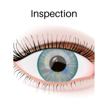
- Check the skin color during skin inspection.
- Check whether redness, cyanosis, pallor, pigmentation, vitiligo, erythema are present in the skin.
- Assess whether any type of rash or lesion is present in the skin.
- If lesion is present, assess its type, size, shape, location, and color.
- In nail examination, check the color, shape, curvature, consistency, surface and clubbing of the nail.
- A pitted surface is seen in the nail in patients with psoriasis.
- Clubbing is seen in people with respiratory diseases.
- In hair inspection, check the hair color, texture, lice, dandruff. In which, check the type of hair, oily, silky, dry, straight, curly.
- Check for conditions such as alopecia and hirsutism.
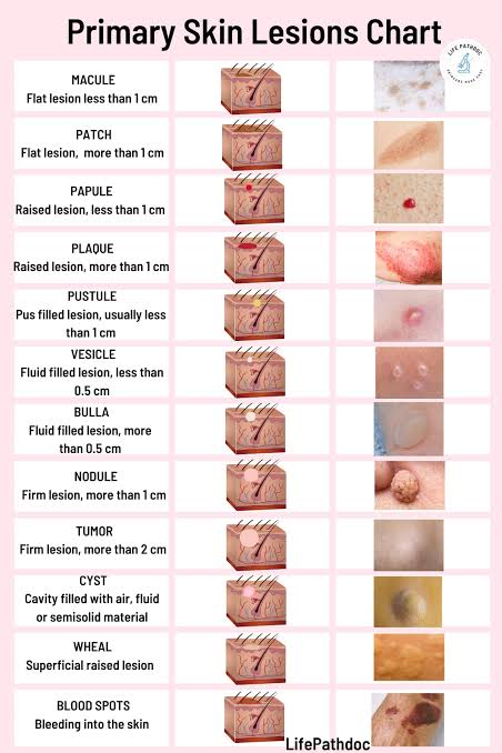
Primary skin lesions
Primary skin lesions are those that occur directly as a result of a disease condition.
1.Macule:-
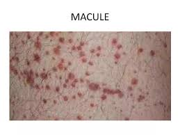
- A macule is flat and non-palpable.
- A macule can be found in different colors, brown, white, purple and red.
- The size of a macule is less than 1 cm.
- Example: Follicles
2. Patch:-
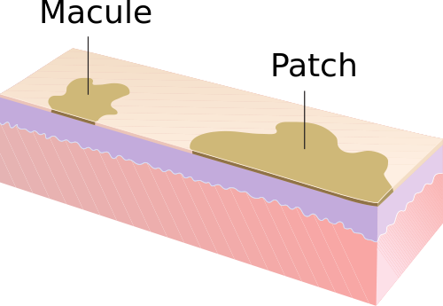
- A patch is a formation similar to a macule (flat and non-palpable) but its size is greater than 1 cm. is.
3. Papule:-
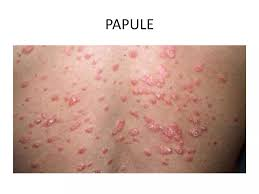
- A papule is an elevated palpable solid mass. Which is less than 1 cm in size. Example: Warts
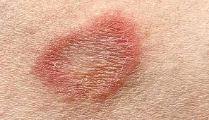
(4) Plaque:-
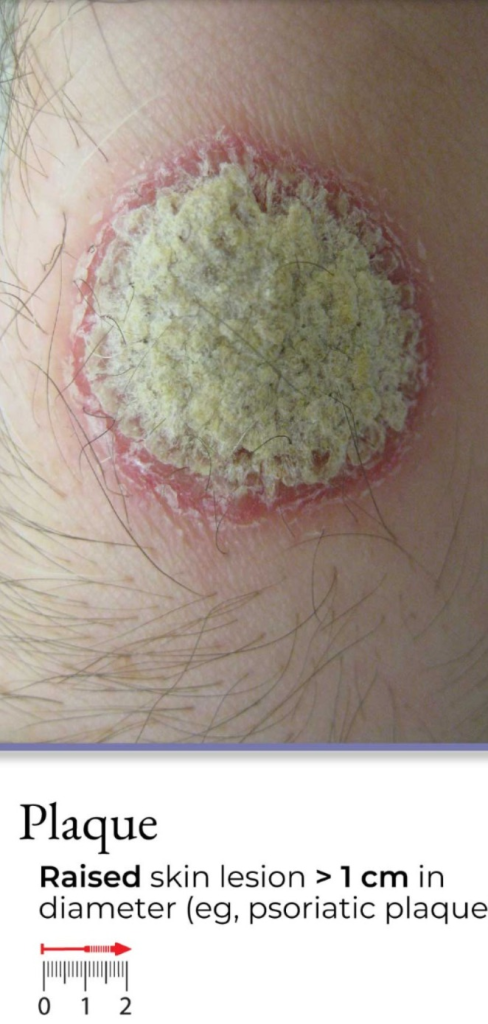
- Plaque is a structure similar to papule (elevated palpable) but its size is more than 1 cm.
- Example: Psoriasis, Keratosis
(5) Nodules:-
- A nodule is an elevated palpable solid mass that extends deep into the dermis layer. The size of a nodule ranges from 0.5 to 2 cm. When the size of the tumor is more than 1-2 cm.
- Example: Lipoma and Carcinoma
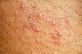
(6) Vesicle:
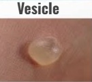
- A vesicle is an elevated palpable fluid-filled mass that is round or oval in shape. Its wall is thin and translucent.
- The size of the vesicle is less than 0.5 cm.
- Example: Herpes simplex, herpes zoster
(7) Bulla (Bulla):-
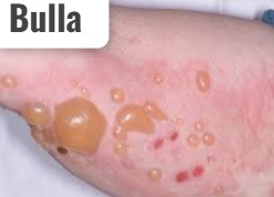
- A bulla is a vesicle-like formation but its size is found to be more than 0.5 cm.
- Example: Pemphigus
(7) Wheal :-
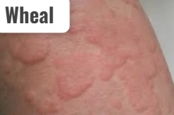
- A wheal is an elevated, radial area with an irregular border.
- Example: UTICARIA, insect bite
(8) Pustule:-
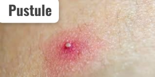
- A pustule is a vesicle containing pus.
- Example: boil, impetigo, furuncle
(9) Cyst (Cyst) :-
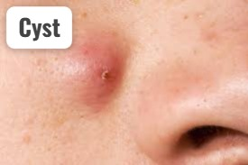
- A cyst is an elevated, fluid-filled semi-solid mass that extends into the subcutaneous tissue and dermis layer.
- Example: Sebaceous cyst
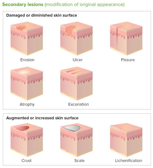
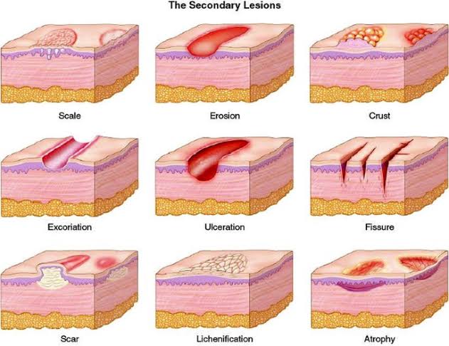
Secondary skin lesions
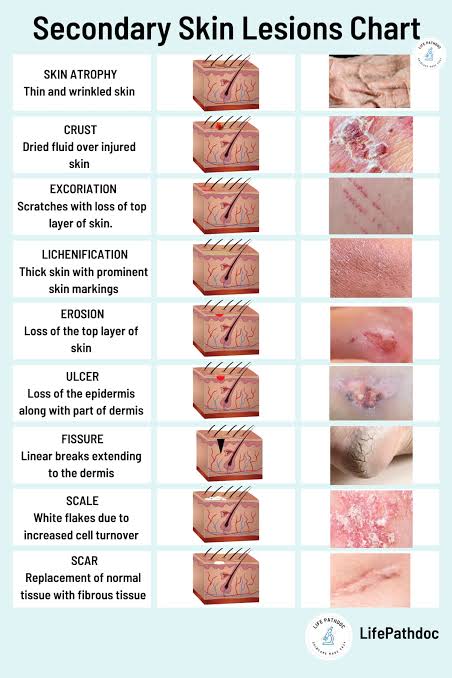
- Secondary skin lesions develop from primary skin lesions. Or it can be seen as a result of etching, infection or trauma.
1) Erosion:
- In erosion, the superficial layer of the skin, the epidermis, is lost or breaks down. Also, that area appears moist and depressed.
- Example: Rupture vesicle and scratch mark
2) Ulcer:
- In an ulcer, the epidermis and dermis layers are affected. That is, deep epidermis layer and necrotic tissue loss are seen
- Example: Pressure ulcer
3) Fissure:
- A fissure is a linear break in the skin. That is, a cut is seen on the skin. Which extends to the dermis layer. Fissures are caused by excessive dryness of the skin and are painful.
- Example: Athlete’s Foot
4) Scales:
- Silver or white flakes appear on the skin due to the accumulation of dead epithelial cells under the skin.
- Example: Dandruff, Psoriasis
5) Scar :
- The mark seen on the skin after a wound or lesion heals is known as a scar. Scars are seen due to the replacement of dead tissue by connective tissue. Young scars are red or purple in color while mature scars are white in color.
- Example: Surgical incision and healing wound
6) Keloid :
- Keloid is an elevated, irregular, red colored hypertrophic scar that occurs due to excessive collagen formation during the healing time.
- Example: Keloid seen on the ear due to surgical incision
7) Atrophy :
- In atrophy, the skin becomes thin, dry and transparent, due to which the vessels underneath it become visible. Which is seen due to loss of collagen and elastin.
- Example: Aged skin and arterial insufficiency
8) Lichenification:
- The skin becomes thick and rough due to repeated, rubbing irritation and scratching.
- Example: Contact dermatitis
9) Crust:
- A crust is a dry exudate (crust) on the skin surface. Which is made up of serum blood and pus.
- Example: Exudate left after rupture of vesicle
Vascular lesion Listen)
1) Petechia :
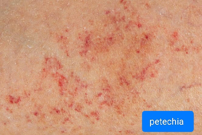
- Petechiae are flat, round-shaped, red or purple spots. They are found to be 1-3 mm in size. Which is seen due to blood leakage in the skin.
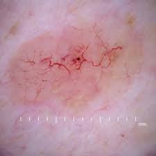
2) Telangiectasia (Telangiectasia):
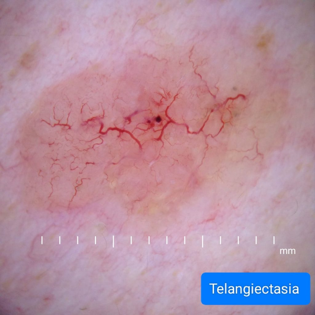
- Telangiectasia is also known as Venus star. Telangiectasia is a spider-like bluish or red colored structure that occurs due to dilation of superficial vessels and capillaries in the skin.
3) Ecchymosis :
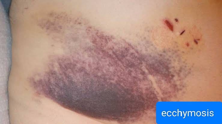
- Echimosis is a macular lesion of round or irregular shape. Which is larger in size than petechiae. In ecchymosis, the skin appears bruised in color due to blood collecting under the skin.
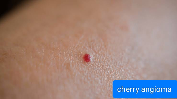
4) Cherry angioma (Cherry angioma):
- A cherangioma is a round-shaped, red or purple colored papule-like structure made up of small blood vessels. Which is seen due to age related skin changes. Which is more common in the trunk and extremities.
5) Spider angioma (Spider angioma):
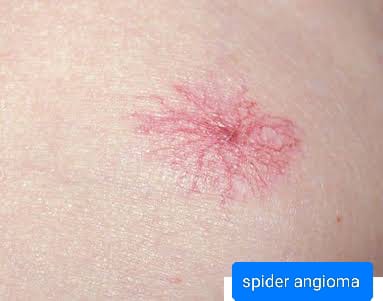
- A spider angioma is a flat vascular lesion of bright red color. Which is seen due to dilation of blood vessels under the skin. Spider angioma is seen in liver disease, vitamin B deficiency and during pregnancy.
Physical Examination :-
Palpation
- Skin temperature, turgor, mobility, moisture, and texture are checked with the help of palpation.
- Check the patient’s body temperature and pulse.
- Palping the skin texture can identify the rashes and lesions in the skin.
- Use gloves while palpating rashes and lesions.
- Before palpating the rashes, gently stretch the skin there so that the redness can be reduced and the rash Can be observed in the same way.
- While palpating the lesion, know its texture, shape and border.
- Palping the lymph nodes in the skin.
- Checking skin turgor and mobility.
- Skin turgor and mobility indicate the elasticity of the skin.
- Skin turgor decreases with age is.
- Screen turgor is seen in dehydrated patients.
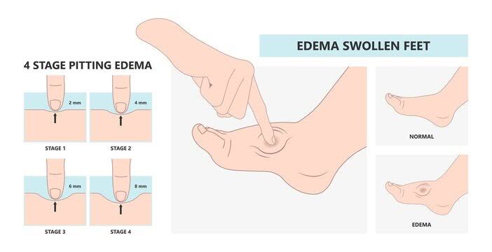
- Finally, check whether edema is present in the legs or not.
- Edema is seen due to excessive fluid accumulation in the body.
- When pressure is applied to this edema, a pit or indentation is seen in it for a long time, it is called pitting edema.
- Pitting edema is commonly seen in the feet and ankles.
Pitting edema can be divided into the following grades.
Grade : 1
- Slight pitting is seen i.e. 2 mm deep pitting is seen and distortion is not seen.
Grade : 2
- Deep pitting is seen i.e. 4 mm deep pitting is seen and distortion is not seen.
Grade : 3
- Pitting is seen up to 6 mm deep and swelling is seen in the extremities.
Grade : 4
- Pitting is seen up to 8 mm deep and distortion is also seen with it.