ENGLISH-UNIT : 1 Oncology : (part-03)
kidney cancer:
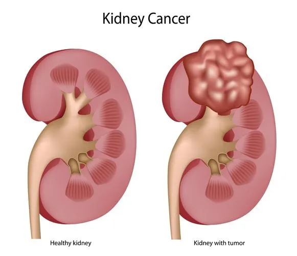
INTRODUCTION ( Introduction ) :
Kidney cancer is also called Renal cancer. In this, abnormal and uncontrolled growth of kidney cells occurs and a tumor forms. These tumors can be both benign and malignant. Kidney cancer mainly arises from two parts of the kidney.
The Renal tubule, ( Renal cell carcinoma),
The Renal pelvis ( transitional cell carcinoma).
If a patient has a renal tumor, he complains of blood in the urine (hematuria) or a mass and pain.
Etiology:
- exact cause is unknown,
- cigarate smoking ,
- Obesity ,
- High blood pressure ,
- long term dialysis,
- occupational exposure to toxic agents,
- certain Analgesic,
- childhood chemotherapy,
- previous radiation therapy .
clinical manifestation :
- Abnormal urin coloure like : dark ,rusty, brown ( Abnormal urine colour : dark, rusty brown ).
- back pain ( back pain ),
- hydronephrosis ( hydronephrosis : accumulation of fluid in the kidney ),
- Abdominal mass or lump ( Abdominal mass or lump ),
- Fever ( fever ),
- Hypertension
- malaise(malaise),
- weight loss(weight loss),
- anorexia (anorexia),
- cold intolerance (Cold Intolerance: Inability to tolerate cold),
- chronic fatigue (chronic fatigue),
- leg and ankle swelling,
- Excessive Night Sweat (excessive night sweat),
- Difficulty seeing,
- hypercalcemia
Diagnostic evaluation:
- history taking and physical examination,
- intra venous urography,
- Cytological examination,
- Renal angiogram,
- ultra sonography,
- ct scan.
Management:
- Radiation therapy,
- Chemotherapy,
- Hormonal therapy.
- Surgical management:
- Nephrectomy
- Simple Nephrectomy: In this, only the tumor is removed.
- Partial Nephrectomy: In this, the tumor and some of the surrounding area are removed.
- Radical Nephrectomy: In this, the kidney, tumor, adrenal gland, lymph node and surrounding tissue are removed.
Nursing management:
preoperative and Postoperative nursing management (Pre-operative and Post-operative Nursing Management) :
Preoperative nursing management (Pre-operative Nursing Management):
- Provide psychological support to the patient.
- Explain the procedure to the patient and his/her relatives.
- Check the patient’s intake output.
- Provide intravenous fluid to the patient.
- Provide blood transfusion to the patient.
- Provide oxygen to the patient.
- Shave the patient on the operative area.
- Provide a comfortable and workable environment for the patient and his/her relatives.
post operative nursing management (Post Operative Nursing Management) :
- Keep the patient under close observation after the operation.
- Check the patient’s vital signs every 15 minutes. Check vital signs.
- Check the patient’s blood pressure every 15 minutes.
- Provide oxygen to the patient if needed.
- Provide intravenous fluid.
- Maintain the patient’s nutritional and hydration status.
- Provide psychological support to the patient and his family members. To do.
- Provide proper antibiotic and analgesia medicine to the patient.
- Properly dress the patient’s operation area.
- Clear all doubts of the patient and his family members.
BLADDER CANCER (Bladder Cancer):
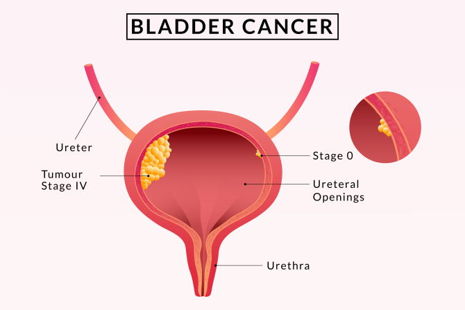
INTRODUCTION (Introduction):
In bladder cancer, there is abnormal and uncontrolled growth of epithelial cells of the bladder and formation of tumors. These tumors can be benign and malignant. Bladder cancer accounts for 90% of all cancers of the urinary system.
The types of cancer in the urinary system are classified according to the cells they form.
urethelial carcinoma ,
squamous cell carcinoma ,
Adenocarcinoma (In adenocarcinoma).
Etiology:
- Age: Mainly seen between the ages of 50 and 70.
- Sex: Affects men more than women. (3 : 1),
- cigarate smoking (cigarette smoking),
- chemical exposure (chemical exposure),
- Diet : It is more common in people who eat fried meat and animal fats.
- Race : Bladder cancer is more common in white people.
- person history of bladder cancer( A person with a history of bladder cancer,
- family history of bladder cancer,
- chronic bladder inflammation,
- Birth defects,
- External beam radiation.
- treatment of certain drugs
clinical manifestation (symptoms and signs):
- blood in urine (hematuria),
- pain during urination,
- frequent urination,
- pelvic pain,
- back pain,
- alteration in voiding.
Diagnostic evaluation:
- history taking and physical examination
- cytoscopy,
- Excretory urography Urography),
- ct scan ( CT scan ),
- ultrasonography ( Ultrasonography ),
- Biannual examination ( Biannual Examination ),
- tumor Biopsy ( Tumor Biopsy ),
- Cytological examination ( Cytological Examination)
medical management ( medical management ):
- Radiation therapy ( radiation therapy),
- chemotherapy ( chemotherapy),
- Immunotherapy ( immunotherapy ).
surgical management:
- cystectomy (removing the bladder)
- partial cystectomy (in which only the affected portion of the bladder is removed).
- Radical cystectomy (in which the entire bladder is removed along with the entire bladder) All surrounding lymph nodes and surrounding tissue structures are removed.
Preoperative nursing management:
In this, the urine output of the patient should be checked every hour in pre-operative and post-operative management.
Properly hospitalize the patient and keep him under close observation of nurses and other health care personnel.
If any complication arises in the patient, immediately inform the health care personnel.
pre operative(pre-operative ):
- Check the patient’s urine output.
- Insert a catheter into the patient.
- Properly explain the surgery, its complications, its benefits and side effects to the patient.
- Check the patient’s vital signs.
- Prepare the patient for surgery.
- Obtain consent for surgery from the patient’s family members.
- Properly explain the removal of the patient’s clothes and jewelry.
- Properly Shave.
- Provide psychological support to the patient and his family members.
- Provide intravenous fluid to the patient.
- Paint the patient’s body area with proper Savlon and spirit.
post operative nursing management (Post Operative Nursing Management) :
- Keep the patient under close observation after the operation.
- Check the patient’s vital signs every 15 minutes.
- Keep a blood transfusion ready for the patient.
- Provide intravenous fluid to the patient.
- Proper dressing of the operative area.
- Provide the patient with proper antibiotic and analgesic medicine.
- Maintain aseptic technique while handling the patient.
- Clear all doubts of the patient and his family members.
- Tell the patient not to do any hard activity.
- Tell the patient to rest completely. Avoid spicy and fatty foods.
- Maintain the patient’s intake output chart.
- Ask the patient to maintain personal hygiene.
- Get all the patient’s blood investigations done.
- Provide psychological support to the patient and his family members.
Ovarian cancer:
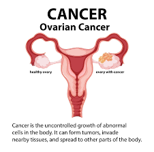
INTRODUCTION ( Introduction):
The ovary is an organ of the reproductive system. Abnormal and uncontrolled growth of its cells in the ovary and the formation of tumor-like structures result in malignancy, i.e. cancer. The result of cancer Bloating ( Tenderness := Pain from intercourse), Pelvic pain, Frequent urination.
Etiology :
- excessive use of birth control pills,
- early menarche (menstrual starts at an early age),
- late menopause
- Nullipara (a woman who has never conceived before the period of pregnancy viability).etc…
clinical manifestation (symptoms and signs):
- Pelvic pain,
- abdominal pain,
- constipation,
- nausea( Nozia),
- weight loss (weight loss),
- poor appetite (poor appetite),
- weakness (weakness),
- Fatige( Fatige).
Diagnostic evaluation
- history tacking and physical examination.
- laparotomy ( laparotomy),
- X ray ( X ray),
- ct scan ( CT scan),
- ultrasound( ultrasound),
- MRI (M. R. I) ,
- to check Elevated serum protein level
- Increase. Ca :=125.
management:
- radiation therapy, ,
- chemotherapy,
- biotherapy,
- surgically remove of tumor ( surgically remove of tumor).
Nursing management (Nursing Management ) :
- Do a head to toe examination of the patient.
- Check the patient’s vital signs To do.
- Check the patient’s intake output.
- See what the patient’s pain level is.
- Check the patient’s skin integrity.
- Maintain the patient’s hygienic condition.
- Provide the patient with a comfortable position.
- Provide Mind Diversion Therapy to the patient.
- Provide Analgesic Medicine to the patient.
- Maintain the hygienic condition of the patient.
- Provide proper bedsheets and clean clothes to the patient.
- Provide Bed Bath and Sponge Bath to the patient.
- The patient Provide clean and wrinkle-free bedsheets.
- Advise the patient to maintain oral hygiene.
- Check the patient’s skin turgor and integrity.
- Instruct the patient to drink two to three liters of water throughout the day.
- Maintain the patient’s nutritional status.
- Provide the patient with a comfortable and functional environment. Providing the environment.
- Providing the patient with proper position.
- Providing proper psychological support to the patient.
- Explain to the patient about the side effects of cancer surgery and chemotherapy and radiation therapy.
- Tell the patient to do some activity every day.
- Explain to the patient that a little exercise every day is necessary. To do.
- Maintain aseptic technique while handling the patient.
- Clear all doubts of the patient and his family members.
- Answer all questions of the patient and his family members. And provide him with psychological support.
cervical cancer:
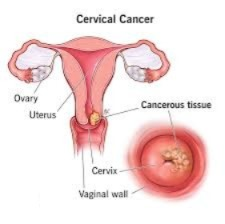
INTRODUCTION ( Introduction ) :
Cervical cancer is the abnormal and uncontrollable growth of cervical cells and the formation of tumor-like structures. These tumors can be benign or malignant.
Etiology (Etiology):
- multiple Sex partners Partner),
- birth control pills (birth control pills),
- Nallipara (Nallipara),
- Multiparty (multiparity),
- human papilloma viral infection(human papilloma virus infection),
- Nutritional deficiency (nutritional deficiency),
- Low socio economic factors,
- early child bearing,
- smoking,
- Chronic cervical infection,
- Hiv infection,
- cigaratte smoking .
clinical manifestation:
- abnormal vaginal bleeding ,
- thin vaginal discharge ,
- Pelvic and low back pain,
- painful urination,
- oedema of lower extremities,
- weight loss,
- anemia
Diagnostic evaluation:
- history tacking and physical examination (history taking and physical examination).
- pap smear test (pap smear test),
- Pelvic examination (pelvic examination),
- X rays ( X rays ),
- laboratory investigation (laboratory investigation),
- Biopsy (biopsy),
- ultrasonography (ultrasonography),
- MRI ( M. R. I. ).
- Calposcopy (calposcopy),
- cytography ( cytography),
- barium X Ray studies (barium X Ray study),
- Intravenous urography.
management:
1) Cryotherapy: In this, the tumor is frozen using liquid nitrogen.
2) (LEEP: Loop Electrocautery Excision Procedure: This procedure is used to remove abnormal cells. In this, a very thin wire is used to make a thin cut in the lesioned layer of the cervix.
3) Conization: In this, a cone-shaped portion of the cervix is removed.
4) Hysterectomy ( Hysterectomy ) : In this, the entire uterus is removed.
5) Total Hysterectomy ( Total Hysterectomy ) : Remove uterus, Cervix, and ovaries.
6) Radical Hysterectomy ( Radical Hysterectomy ) : Remove uterus, Cervix,ovaries,Fallopian tube, Malignant area of vagina affected lymph node.
7) Pelvic trachelectomy ( Pelvic trachelectomy): In this, only the selected part of the cervix is removed. And at the same time, the tumor in the cervix is removed.
8) Radiation therapy, ,
9) Chemotherapy, .
preoperative and Postoperative nursing management:
preoprative nursing management (Preoperative Nursing Management) :
- Provide psychological support to the patient.
- Explain the entire surgical procedure to the patient.
- Prepare the patient physically and mentally for surgery.
- Maintain the patient’s nutritional status.
- Maintain the patient’s hydration status.
- Catheterize the patient.
- Inject the patient with I.v. Set up the line.
- Provide fluids to the patient through the parenteral route.
- Perform all laboratory investigations on the patient.
- Provide a comfortable and working environment for the patient.
- Keep blood transfusions ready for the patient.
- Properly shave the operative area of the patient’s body To do.
- Paint the patient’s operative body parts with proper Savlon and spirit.
- Give the patient the prescribed antibiotic medicine in the proper manner.
- Check the patient’s vital signs.
post operative nursing management (Post Operative Nursing Management) :
- Close observation of the patient after the operation.
- Maintain aseptic technique while attending to the patient.
- Check the patient’s vital signs every 15 minutes.
- Proper dressing of the patient’s operative area.
- Proper observation of the surgical site Maintain the patient’s intake output chart. Provide intravenous fluid to the patient. Provide the patient with prescribed analgesic and antibiotic medicine. Do not check the drainage tube properly. Instruct the patient to monitor the operative site for any redness, swelling, To see if there is inflammation.
- Provide psychological support to the patient and his family members.
- Encourage the patient to do hard activities.
- Instruct the patient to do small and frequent amounts of activity.
uterine cancer ( uterine cancer ):
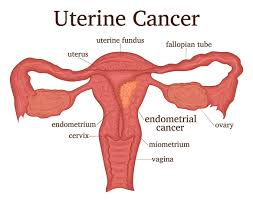
The uterus is an organ of the female reproductive system.The uterus is an organ of the female reproductive system.The abnormal growth of uterine cells in the uterus And due to uncontrollable growth, a tumor is formed. This tumor can be benign (noncancerous) or malignant (cancerous) and it mainly occurs in the endometrium (innermost layer) of the uterus.
Etiology:
- chronic exposure to estrogen (long-term exposure to estrogen).
- Endometrium Hyperplacia (endometrium hyperplasia),
- Obesity (obesity),
- a high fat diet (high fat food),
- diabetes (diabetes),
- Women over 50 -60 years of age.
- multiparty( multiparity ),
- nallipara ( nalipara ),
- Hiv infection (HIV infection),
- nutritional deficiency ( nutritional deficiency ).
- family history( in a family with a history),
- exposure to radiation( radiation) contact, )
- race (white women’s are more likely to have uterine cancer).
clinical manifestation (symptoms and signs ):
- abnormal vaginal bleeding (vaginal discharge),
- painful and difficulty in urination (painful urination),
- Pelvic pain (pelvic pain),
- anemia (anemia),
- Fatige (fatigue),
- weakness (weakness).
Diagnostic evaluation:
- History tacking and physical examination,
- Biopsy,
- ultrasound
- X Ray,
- Computed Tomography (Ct scan),
- Magnetic Resonance Imaging (MRI)
- transabdominal ultrasound,
- transvaginal ultrasound
Management:
- Radiation therapy,
- Chemotherapy,
- hormone therapy,
- If the tumor is small, conservative treatment should be provided.
- Use gonadotropin releasing hormone analogs to reduce estrogen hormone levels.
- Obesity of the patient Level control.
surgical management:
- if large tumor so do myomectomycc
- laproscopic myomectomy
- hysterectomy( Hysterectomy),
- partial hysterectomy,
- total hysterectomy.
preoperative and Postoperative nursing management:
Preoperative nursing management:
- Provide complete information about the treatment and disease to the patient.
- Explain all the pre-operative and post-operative procedures to the patient.
- Check the patient’s vital signs.
- To Provide analgesic and antibiotic medicine.
- Provide the patient with all the information about the operation and obtain consent from the patient’s family members.
- Prepare the patient physically and psychologically for the operation.
- Set up two intravenous lines for the patient.
- Perform all laboratory investigations on the patient.
- Keep blood ready for the patient.
- Provide intravenous fluid to the patient.
- Shave the patient’s body parts where the operation is to be performed properly.
- Clean the patient’s body parts with Savlon and Betadine.
- Insert a urinary catheter into the patient.
- Maintain the patient’s intake output chart.
- Maintain aseptic technique while handling the patient.
- Paint the patient’s operative area with proper Savlon and Betadine.
post operative nursing management (Post Operative Nursing Management) :
- Keep the patient under close observation after the operation.
- Watch the patient for any complications after the operation.
- Check the patient’s vital signs every 15 minutes.
- Maintain the patient’s intake output chart.
- Provide intravenous fluid to the patient.
- Administer blood if the patient has lost an excessive amount of blood.
- Properly dress the patient’s operative parts.
- Maintain aseptic technique while handling the patient.
- Provide the patient with prescribed analgesic and antibiotic medicine.
- Change the patient’s position frequently to prevent bed sores.
- Check the patient’s vital signs.
- Maintain aseptic technique while handling the patient.
- Provide psychological support to the patient and his family members.
- Tell the patient and his family members to first introduce the patient to a liquid diet, then a semi-solid, and then a solid diet.
- Tell the patient not to do any kind of hard work.
- Tell the patient to move around a little.
- Educate the patient to take proper bed rest.
- Proper follow up with the patient and his family members to say.
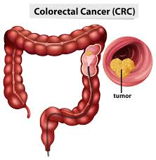
colorectal cancer:
Introduction (Introduction):
COLON is an organ of the gastrointestinal system. Abnormal and uncontrolled growth of cells in the colon leads to the formation of tumors and these tumors can be both benign and malignant. In colorectal cancer, cancerous growths occur in the colon, rectum, and appendix and affect the digestion of food.
Etiology:
- age : above 50 year old age,
- Diet( डियाट ),
- Genetic disorder ( गेनेटिक दीश्वर्डर ),
- Family history( फिमी हिस्ट्री ),
- personal history of polyps ( प्रसनल हिस्ट्री अफ पोलिप्स ),
- history of Inflammatory bowel disease (History of Inflammatory Bowel Disease),
- Obesity( Obesity),
- viral( Virus),
- smoking( Smoking),
- alcohol( Alcohol),
- excessive use of fatty and spicy food,
- male are more affected than female,
- excessive use of fat.
clinical manifestation (symptoms and signs):
- Fatigue,
- Weakness,
- Difficulty in Breathing,
- Changes in Bowel Habit.
- small – caliber or ribbon like stool,
- Diarrhea,
- red and dark blood in stool,
- nausea,
- vomiting,
- weight loss,
- rectal pain,
- abdominal pain,
- Distention,
- cramp
- Bloating
Diagnosis evaluation:
- History taking and physical examination,
- stool test,
- fecal occult blood test,
- colonoscopy Colonoscopy),
- Genetic testing.
Management:
- Radiation therapy,
- chemotherapy,
- biotherapy,
- Genetherapy,
- immuno therapy.
Surgery:
- Surgery is the choice for colorectal cancer.
- Radical bowel resection,
- partial colostomy,
- hemicolectomy,
- laproscopic surgery.
Prevention:
- Regular screening,
- Genetic counselling,
- Lifestyle and nutrition,
- Quit smoking.
Preoperative and Postoperative nursing management:
Preoperative nursing management:
- Explain the procedure to the patient and his family members.
- Get all the laboratory investigations done on the patient.
- Prepare the patient for the operation.
- Set up the IV line on the patient.
- Keep the patient on NBM (nill per oral).
- Catheterize the patient.
- Maintain the patient’s intake output chart.
- Shaving the patient’s operative body parts properly.
- Clean the patient’s operative body parts with Savlon and spirit.
- Provide the patient with eye fluid.
- Provide the patient with total parenteral nutrition.
- Maintain the patient’s nutritional status and food balance normal.
- Provide the patient with prescribed analgesic and antibiotic medicine.
- Obtain the consent of the patient and his family members.
Post operative nursing management:
- Keep the patient comfortable and under close observation after the operation.
- Provide the patient with a comfortable and workable environment.
- Ask the patient to do deep breathing exercises.
- Keep the patient’s fluid balance normal.
- Maintain the patient’s aseptic technique.
- Maintain the patient’s nutritional status.
- Maintain the patient’s intake output chart.
- Dress the patient’s operative area properly.
- Check the patient’s operative area for any infection or inflammation.
- Check the patient for any weakness or nausea and vomiting.
- Check the patient’s vital signs.
- Provide the patient with I.v. fluids.
- Change the patient’s dressing every twenty-four hours.
- Provide the patient with prescribed analgesic and antibiotic medicines.
- Maintain hygienic condition of the patient.
- Change the patient’s position every two hours to prevent bed sores.
- Ask the patient to move around a little.
- Do not ask the patient to do any kind of hard activity.
- Ask the patient to take proper rest.
- Provide mind diversion therapy to the patient.
- Provide liquid, semi-solid and then solid food to the patient first.
- Keep the patient’s head elevated to prevent any heart burn.
- Provide psychological support to the patient and his family members.
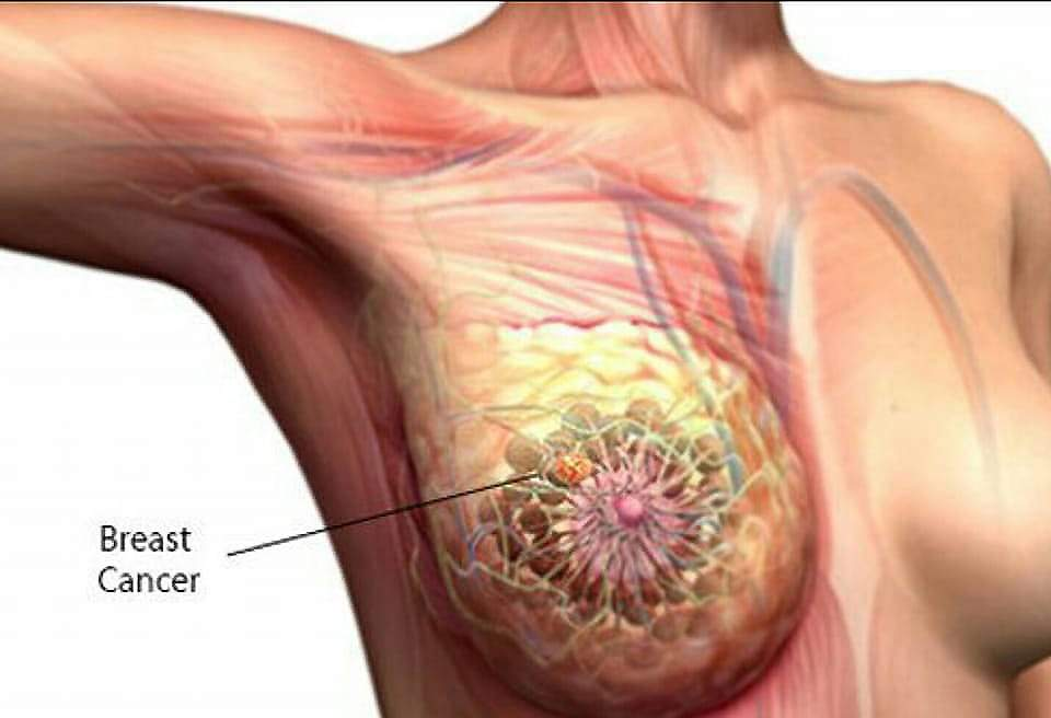
Breast cancer:
Introduction :
Breast cancer is the abnormal and uncontrolled growth of breast cells, which results in a tumor, which can be malignant. It is called breast cancer. Breast cancer can occur anywhere in the breast, but it is most common in the upper outer parts of the breast, where there is more tissue. Breast cancer arises from cells in the lobules of the breast, which are called milk-producing glands or ducts.
Etiology:
- age: in women over 60 year old age
- Gender: most in female
- personal history of breast cancer
- family history
- certain breast change
- reproductive and menstrual history
- atypical Hyperplacia
- Hormonal replacement therapy (estrogen and progesterone),
- oral contraceptive use,
- overweight and Obesity,
- lack of physical activity,
- drinking alcohol.
- a lump or thickening in or near the breast or in the underarm area.
- There is a change in the size and shape of the breast.
- The skin of the breast areola and nipple becomes red, swollen and scaly.
- Irritation and dimpling of the skin.
- Pain in the breast.
- Pain and tenderness in the nipple.
- The nipple turns inward in the breast.
- Discharge comes out of the nipple.
- Axillary and The supraclavicular lymph node enlarges.
- History taking and physical examination
- Biopsy,
- X Ray (X-ray),
- ct scan (CT scan),
- MRI (M. R. I.),
- mammography (Mammography).
- Radiation therapy,
- Chemotherapy,
- Biotherapy,
- Gene therapy.
clinical manifestation (symptoms and signs):
Diagnostic evaluation:
Management (Management):
surgery:
- Mastectomy,
- partial Mastectomy,
- Radical Mastectomy,
- lymph node dissection,
- cryotherapy,
- breast reconstructive surgery.
preoperative and Postoperative nursing management:
Preoperative nursing management:
- Close observation of the patient to see if there are any side effects of radiation therapy such as fatigue, sore throat, cough, nausea, loss of appetite.
- To see if the patient has any side effects of chemotherapy such as bone marrow Suppression, hair loss, weight loss, fatigue, depression, anxiety, etc. should be checked.
- Provide psychological support to the woman.
- Involve the woman in the treatment.
- Explain all the procedures to the woman.
- Provide antiemetic medicine to the patient.
- Setting up an IV line for the patient.
- Providing IV fluid to the patient.
- Catheterizing the patient.
- Maintaining the patient’s intake output chart.
- Preparing the patient’s body part for surgery in a proper manner.
- All laboratory investigations of the patient To be done.
- Obtain the consent of the patient and his family members.
post operative nursing management (Post Operative Nursing Management) :
- Keep the woman under close observation after the operation.
- Check the patient’s vital signs every 15 minutes.
- Check the blood pressure every 15 minutes.
- Provide psychological support to the patient Do.
- Provide the patient with a comfortable and functional environment.
- Give the patient an I.V. Provide fluid.
- Maintain the patient’s intake output chart.
- Properly dress the patient’s operative body parts.
- Provide the patient with a comfortable and working environment.
- Check the patient’s body part where the surgery was performed for any redness, swelling, or inflammation.
- Provide antibiotic and analgesic medicine to the patient.
- Maintain the nutritional and hydration status of the patient.
- Provide the patient with his/her JC and antibiotic medicine.
- Ask the patient to do some exercise.
- Provide psychological support to the patient and his/her family members.
- Ask the patient to maintain hygienic conditions.
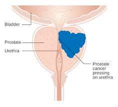
Prostate cancer:
Introduction:
Prostate cancer occurs in the prostate gland. In the prostate gland, there is abnormal and uncontrolled growth of prostate gland cells and formation of tumors and this tumor results in a cancerous tumor and causes cancer . Prostate cancer can also spread to surrounding tissues and this cancer can spread to all parts of the body such as liver, bone, lungs etc.
Etiology:
- advance age
- hereditary,
- Hormonal influence ( Hormonal influence ),
- environment Factor ( Environmental Factor ),
- cigarate smocking ( Cigarette Smoking ),
- toxins, chemical( Toxins Chemical).
- industrial products( Industrial Product ).
- diet high in saturated fat
clinical manifestation (symptoms and signs ):
- burning or pain during urination,
- inability to urinate ),
- a sensation of incomplete emptying of the bladder even after passing urination in urine (blood present in urine),
- Pelvic pain(pelvic pain),
- back or hip pain( back and hip pain),
- abdominal pain( abdominal pain),
- chest pain( chest pain),
- weight loss( weight loss).
Diagnosis evaluation ( Diagnostic evaluation ):
History taking and physical examination,
Biopsy,
blood test (blood test),
urine test (urine test),
X Ray (X Ray),
ct scan (Computed Tomography),
MRI (Magnetic Resonance Imaging),
Management (Management):
-
- Radiation therapy,
-
- Chemotherapy,
-
- Biotherapy
-
- gene therapy,
-
- immuno therapy,
-
- Hormonal therapy,
-
- cryotherapy,
Nursing management:
assessment:
-
- Perform a head to toe examination of the patient.
-
- Check the patient’s vital signs.
-
- Maintain the patient’s intake output chart. Assess the patient’s pain level.
-
- Assess the patient’s skin integrity.
-
- Assess the patient’s bowel and bladder habits. To do an asaess.
-
- Maintain the patient’s oral hygiene.
-
- To see if the patient has hair loss or any side effects due to chemotherapy and radiation therapy.
Nursing diagnosis
1) pain related to disease condition
Nursing interventions:
Reliving pain level:
-
- Assess the patient’s pain level.
-
- Provide the patient with a comfortable position.
-
- Provide the patient with mind diversionary therapy.
-
- The patient Provide analgesics.
-
- Provide a comfortable and functional environment for the patient.
2) Self care deficit related to disease condition.
Nursing interventions:
provide hygiene conditions to the patient:
-
- Provide the patient with clean clothes and bed sheets.
-
- Provide the patient with bed bath and sponge bath.
-
- Provide the patient with clean and wrinkle-free bed sheets.
-
- Maintain oral care for the patient.
-
- Instruct the patient to wash their hands properly.
3)Impaired skin integrity related to cancerous condition.
Nursing interventions:
mainten skin integrity.
- Assess the patient’s skin integrity.
- Check the patient’s skin turgor.
- Check whether the patient is bleeding from the body.
- Give the patient two to three liters of Ask the patient to drink water.
- Ask the patient to apply lotion to the body.
- Tell the patient not to rub the skin and not to scratch it.
- Tell the patient to wear cotton clothes.
- Do not allow the patient to wear tight clothes.
4)Impaired nutrition status of patient less than body requirement related to diarrhea and vomiting .
Nursing interventions:
improve nutritional status:
- Nutritional status of the patient Assess.
- Provide a comfortable environment for the patient.
- Ask the patient to wash their hands while eating.
- Provide the patient with an appetizer.
- Remove any items in bad order from the patient’s surroundings while they are eating.
- Consider the patient’s likes and dislikes Keep.
- Feed the patient at regular intervals.
- Give the patient only the food he likes.
- When the patient is eating, his environment should be completely keep clean.
5) Disturbed body image (hair loss) related to radiotherapy and chemotherapy.
Nursing interventions:
improve body image of patient:
- Maintain good interpersonal relationship with patient.
- Maintain good rapport with patient.
- Tell patient that hair loss is temporary.
- Tell patient that when chemotherapy and Hair loss is common while undergoing radiation therapy.
- Provide psychological support to the patient.
6)activity intolerance related to the weakness.
Nursing interventions:
improve the activities of the client.
- Assess the patient’s activity level.
- Maintain a good interpersonal relationship with the patient.
- Instruct the patient to do daily routine activities in small increments.
- Instruct the patient to Take some rest between activities.
- Tell the patient to do proper exercises and yoga.
7)High risk of infection related to *hospitalizations .
Nursing interventions:
reduce the risk of infection:
- Assess the patient’s infection side.
- Maintain aseptic technique while handling the patient.
- Dressing the patient’s body parts Maintain aseptic technique while performing the procedure.
- Provide proper clean and hygienic conditions to the patient.
- Give the patient clean clothes to wear.
- Give the patient high protein rich food that helps the patient fight infection.
- Provide the patient with proper antibiotic medicine.
8) depression and fear related to treatment of cancer .
Nursing interventions:
Reduce the fear level of client.
- Check the patient’s anxiety and fear level.
- Maintain a good interpersonal relationship with the patient.
- Help the patient to solve his doubts and problems.
- Listen properly to the patient and his family members.
- Answer all the patient’s questions.
- Provide psychological support to the patient.
- Provide coping skills on how the patient can deal with such a bad situation.
Bone cancer:
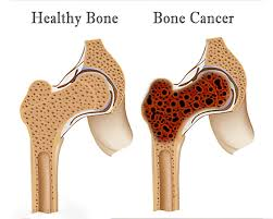
INTRODUCTION ( Introduction ) :
Bone cancer can occur in any part of the body. But bone cancer mainly affects the long bones, such as the bones of the arms and legs. If the cancer originates in the bone, it is called primary bone cancer, and if the bone cancer spreads from another part of the body and spreads to the bone, it is called secondary bone cancer.
type of bone cancer( Type of bone cancer ):
1)benign bone tumor ( Benign bone tumor):
Benign bone tumor include:
- osteomas ( osteomas),
- osteoblastomas ( Osteoblastomas),
- osteoidosteoma ( osteoid osteoma),
- osteochondromas ( osteochondromas),
- enchondroma ( enchondroma)
2)malignant bone tumor:
- The most common bone tumor
- osteosarcoma ( osteosarcoma),
- chondrosarcoma ( chondrosarcoma),
- fibrosarcoma ( fibrosarcoma),
- chordoma ( chordoma)
3)Metastasis bone cancer :
- Almost all types of cancer spread to the bone but mainly
- Bone,
- Breast,
- Lungs,
- Kindny,
- Thyroid And Prostate ( Thyroid And Prostate )
- These are the main Organ from which cancer is transmitted to the bone.
Etiology:
- occurs at 10-25year age,
- exposure to radiation,
- inherited genetic disorder( in Inherited genetic disorder),
- some drugs,
clinical manifestation( symptoms and signs ):
- pain,
- mass or lump felt in the bone( lump or mass felt in the bone) structure filling),
- weak bone,
- fever,
- chills,
- night sweat,
- anemia,
- anorexia,
- fatigue,
- Tenderness,
- weight loss,
- neurological symptoms may present with nerve root compression ( neurological symptoms may also be seen).
- Swelling ,
- limited motion ( limited motion ),
- increase skin temperature over mass ( increase skin temperature over mass ).
Diagnostic evaluation:
- History tacking and physical examination,
- bone scan,
- X Ray (X-ray),
- ct scan (CT scan),
- MRI (M. R. I.),
- myelography (myelography),
- arteriography (arteriography),
- Biopsy (biopsy),
- Elevated syrum alkaline Phosphate (elevated serum alkaline phosphate).
Management:
- Radiation therapy,
- Chemotherapy,
- Biotherapy
- Bone Merrow transplantation
- Immunotherapy,
- Immunotherapy,
- Gene therapy.
surgical management:
- limb sparing surgery,
- amputation,
- lymph node dissection node dissection),
- Reconstructive surgery ,
- tumor curettage,
- bone grafting,
- limb salving procedures.
Nursing management:
- To check the patient’s vital signs.
- To see how much blood the patient has lost.
- To see if the patient has any complications. like deep vein thrombosis, Pulmonary embolism, Infection, Etc.
- Give analgesic medication to the patient.
- Provide intravenous fluid to the patient.
- Provide support to the affected extremity with pillows.
- Provide splint to the patient for additional protection.
- Patient and his Ask family members to discuss their fears and ask all their questions.
- Provide psychological support to the patient.
- Prepare the patient in advance for the patient’s body image to change.
- Encourage the patient to do their daily routine activities.
- Provide the patient and his family members with complete information about the disease and its treatment.
- Maintain the patient’s nutritional and hydration status.
- Provide a comfortable environment and work to improve the patient’s sleep pattern.
- Ask the patient to move the affected extremities.
- Provide support to the patient’s joints.
- Be careful not to let the patient fall.
- Create a hazard-free environment for the patient.
- Provide a supportive environment for the patient.
- Provide psychological support to the patient.
- Answer all the patient’s questions.
- Provide psychological support to the patient to improve the patient’s coping ability.
oncological emergency:

This is a life-threatening condition that requires immediate management.
1) superior vena cava syndrome ,
2) hypercalcemia,
3) Spinal cord compression,
4) Pericardial effusion,
5) Intra vascular Coogulopathy,
6) tumor lysis syndrome,
7) Inappropriate antiduretic hormone.
1) superior vena cava syndrome (Superior Vena Cava Syndrome)
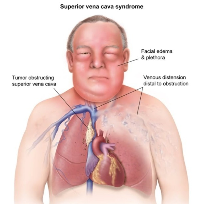
- Superior vena cava syndrome is characterized by obstruction of the superior vena cava, which is mainly seen in cancerous patients. It can cause partial or complete obstruction, which blocks the blood flowing from the head, neck, and upper limbs to the right atrium through the superior vena cava. In this, the superior vena cava is compressed, which can lead to Due to cancerous tumors, enlargement of the lymph nodes causes compression of the superior vena cava and reduces venous circulation or drainage of the head, neck, arm, and thorax. Etiology: Cigarette smoking, tobacco use ),
Etiology :
- cigarette smoking
- non neoplastic disease such as tuberculosis,
- Chronic bronchitis,
- Emphysema.
clinical manifestation (Symptoms and Signs):
- Cough,
- Difficulty in breathing,
- Chest Pain,
- Excessive phlegm,
- Blood in the phlegm Bleeding),
- Fever,
- Nausea and Vomiting,
- Weight loss,
- Fatigue,
- Loss of appetite,
- persistence pneumonitis.
Diagnosis evaluation ( Diagnostic evaluation ):
- History tacking and physical examination,
- X Ray ( X Ray ) ,
- ct scan ( computed tomography) ,
- pet ( positron emission tomography),
- Cytological examination
- Bronchoscopy
- lymph node Biopsy
- PFT (Pulmonary function test).
Management:
- Radiation therapy,
- chemotherapy,
- biotherapy,
- bone marrow therapy
- immuno therapy,
- surgical resection( surgical resection ).
complications ( complications ):
- hypocalcemia ( hypocalcemia ),
- syndrome of inappropriate antiduretic hormone (syndrome of inappropriate antidiuretic hormone),
- spinal cord compression( spinal cord compression),
- pulmonary scaring( pulmonary scaring).
Nursing management (Nursing management):
Nursing assessment :
- Assess the patient’s respiratory rate and vital signs.
- Assess the patient’s urine output.
- Assess the patient’s pain level.
- Assess the patient’s eating pattern.
- The patient’s anxiety level and coping skills To assess.
- To assess the patient’s blood investigations.
- To observe the patient’s signs and symptoms and perform a physical examination.
Nursing diagnosis (Nursing Diagnosis) :
1)Impaired breathing pattern related to compromised respiration.
Nursing interventions:
improve breathing pattern:
- Elevate the patient’s head so that he can breathe properly.
- To give the patient breathing exercises Say.
- Give the patient the prescribed treatment and administer antimicrobial agents and nebulization.
- Give the patient adequate oxygen.
- Sit the patient on a chair and position him in such a way that he can breathe properly.
2)Impaired nutrition pattern less than body requirement related to nausea and vomiting.
Nursing interventions:
improve nutritional status:
- Tell the patient to eat small amounts of high-calorie and high-protein food.
- How to ensure that the patient gets enough protein such as milk, eggs, chicken, fish, cheese, etc.
- Ensure that the patient gets enough Give vitamins in moderation.
- Give the patient a food he likes.
- Provide total parental nutrition to the patient.
3)pain related to abnormal cell growth.
Nursing interventions:
controlling pain:
- Assess the patient’s pain level.
- Give analgesic medications to the patient.
- Provide mind diversionary therapy to the patient.
- Providing reassurance to the patient.
4) Anxiety, fear related to therapeutic regimen and prognosis.
Nursing interventions:
Minimizing anxiety:
- Assess the patient’s anxiety level.
- Ask the patient to explain his/her feelings.
- Provide the patient with NSAID (non steroidal anti inflammatory drugs) drugs.
- Explain the procedure to the patient.
- Maintain good communication with the patient.
Spinal cord compression ( Spinal cord compression):
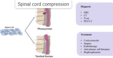
Introduction:
Spinal cord compression occurs due to enlargement of the lymph nodes and formation of tumors, which causes compression of the spinal cord and affects the nervous system. Nervous system abnormalities occur. This abnormality leads to morbidity and mortality.
Etiology:
- tumor,
- lymphomas,
- intervertebral collapse ,
- Metastasis cancer ( Metastasis cancer )(Breast,lung,myeloma Lymphoma.).
clinical manifestation( Symptoms and signs ):
- local inflammation( Local inflammation ),
- Swelling,
- venous stasis,
- Impaired blood supply,
- Pain,
- neurological Dysfunction,
- weakness,
- bladder and bowel Dysfunction.
Diagnostic evaluation:
- History tacking and physical examination (History tacking and physical examination),
- percussion Tenderness at the level of compression(Percutaneous Tenderness at the level of compression).
- abnormal reflexes(Abnormal reflexes),
- sensory and motor abnormality(Sensory and Motor Abnormalities),
- X Ray (X-ray),
- ct scan (CT scan),
- MRI (M. R. I.).
Management (Management):
- Radiation therapy (Radiation therapy) therapy),
- Chemotherapy,
- surgery,
- Biotherapy
- bone marrow therapy
- Gene therapy.
Nursing management:
- Proper assessment of the patient.
- Neurological assessment of the patient.
- Controlling pain levels.
- Preventing the patient from complications.
- Ask the patient to exercise.
- Check the patient’s bowel and bladder habits.
- Reassure the patient.
- Ask the patient and his family members for coping abilities.
- Provide psychological support to the patient and his family members.
hypercalcemia:
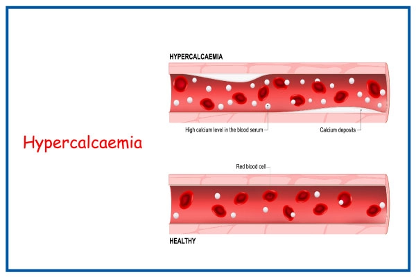
Introduction:
In hypercalcemia, the calcium level in the body increases above the normal level. The calcium level in the body increases due to kidney cancer. Due to kidney cancer, the kidneys do not work properly and due to this, the calcium level in the body increases and due to this, the calcium level in the body increases and it results in hypercalcemia.
Etiology:
bone bone destruction by tumor cell,
excessive use of calcium,
inadequate absorption of calcium in the body.
Diagnostic evaluation:
serum calcium level exceeding 11mg/dl ( Serum calcium level more than 11mg/dl. )
Management:
- Provide proper fluids to the patient.
- Provide diuretic medicine to the patient.
- Use calcitonin to lower calcium levels.
- Identify patients who are at risk of developing hypercalcemia.
- Provide education to patients and family members To do.
- Explain to the patient that dietary and pharmacological interventions such as stool softeners and laxatives should be used.
- Tell the patient to maintain his nutritional status.
- Provide antiemetic medicine if the patient is nauseated and vomiting.
- Instruct the patient to move around a little.
pericardial effusion:
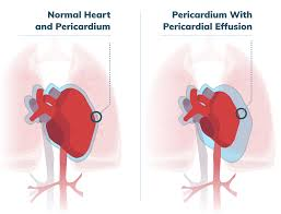
Introduction:
In this, fluid collects in the pericardial (the topmost membrane of the heart) due to lung and esophagus cancer. It is called pericardial effusion.
Etiology:
- cancerous tumor ( cancerous tumor ),
- tumor of lung and Esophagus,
- breast cancer.
clinical manifestation:
- compensatory tachycardia( Compensatory tachycardia),
- increaed vascular pressure( Increase vascular pressure),
- narrow pulse pressure( Narrow pulse pressure),
- lowest blood pressure( Lowest blood pressure),
- shortness of breath( Shortness of breath),
- weakness( Weakness),
- chest pain,
- anxiety,
- lethargy.
Diagnostic evaluation:
- History tacking and physical examination (History Taking and Physical Examination),
- E.C.G.( E.C.G. ),
- chest x-ray( Chest X-ray),,
- ultrasound( Ultrasound )
- ct scan (CT Scan),
- MRI (m. r. i.).
Management:
- pericardial aspiration.
- Closely monitor ECG.
- Instruct cardiologist while performing pericardial aspiration.
- Carefully monitor cardio vascular pressure.
- Maintain hygienic condition of patient.
- Maintain the nutritional status of the patient.
- Give reassurance to the patient.
- Provide a comfortable position to the patient.
- Provide comfort to the patient.
- Monitor the patient for any other complications.
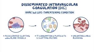
Disseminate intravascular coagulopathy:
Introduction:
Disseminated intravascular coagulation (DIC) is a serious condition where the body’s blood clotting system becomes overactive and widespread, leading to abnormal blood clotting and excessive bleeding. It is not a disease in itself, but a complication of other underlying conditions. DIC involves systemic activation of coagulation, leading to microvascular thrombosis (small blood clots) and potentially life-threatening hemorrhage due to the consumption of platelets and clotting factors. This bleeding can occur both internally and externally.
Etiology:
- Haematological Cancer,
- Prostate Gland Cancer,
- Abnormality in Gastrointestinal track and Lungs.
- Chemotherapy,
- sepsis,
- Hepatic failure (hepatic failure).
Diagnostic evaluation:
- History tacking and physical examination,
- decrease hemoglobin,
- decrease hematocrit Hematocrit),
- Elevated fibrinogen split products,
- positive protamine sulfate precipitation test.
clinical manifestation( symptoms and signs ):
- blood clot formation,
- greater risk for Impaired circulation,
- tissue hypoxia,
- fibrinolysis may occur.
Management:
- Check the patient’s vital signs.
- Check the patient’s intake output.
- Check the patient’s skin integrity.
- Check the patient’s vital signs and especially temperature.
- Check the patient’s visual acuity.
- Check if the patient has chest pain.
- Check if the patient has abdominal tenderness.
- Check the patient’s body orifices through which the tube was inserted to see if there is any bleeding.
- Perform laboratory tests on the patient.
- Assess the patient’s oxygen level.
- Apply more pressure to the site where the patient’s vein was punctured.
- Maintain the patient’s oral hygiene.
- Teach the patient deep breathing exercises.
- Provide the patient with a comfortable and functional environment.
Syndrome of inappropriate secretion of antidiuretic hormone (SIADH):

Introduction:
Syndrome of Inappropriate Antidiuretic Hormone (SIADH) This is a condition in which the uncontrolled release of antidiuretic hormone (ADH) from an abnormal tumor or due to abnormal stimulation of the hypothalamic pituitary network occurs and due to this, the volume of extracellular fluid increases and a condition of hyponatremia (low level of sodium in body) occurs.
Etiology:
- cancer of lungs (lung cancer),
- Antineoplastic agents also stimulate ADH (antidiuretichormone) secretion.
clinical manifestation (symptoms and signs ):
- A person’s personality changes.
- irritability Occurs.
- Vomiting (vomiting),
- Increase Weight (weight gain),
- Head Ache (headache),
- Fatigue (fatigue),
- confusion (confusion),
- seizure ( hesitating),
- abnormal reflexes ( Abnormal reflexes ),
- papilledema ( Papilloedema ),
- coma ( Coma ),
- death ( Death ).
Diagnostic evaluation (Diagnostic Evaluation):
- History tacking and physical examination (History Taking and Physical Examination),
- decrease Cirum sodium level( Decrease Serum Sodium Level),
- increase urine osmolarity( Increase Urine Osmolarity),
- decrease creatinine And Albumin (Decreases creatinine and albumin).
Management:
- Reduce fluid intake to 500 to 1000 ml/day Intake fluid.
- Use diuretic therapy.
- Monitor the patient’s intake output.
- Check the patient’s level of consciousness.
- Listen to the patient’s lungs and heart sounds.
- Check the patient’s vital signs. –>
- Check the patient’s weight daily.
- Check the patient’s urine specific gravity.
- Get the patient’s laboratory tests done.
- Check the patient’s electrolytes.
- Get the patient’s blood and urine tests done.
- Reduce the patient’s activity level.
- Maintain the patient’s oral hygiene.
- Maintain the patient’s environmental safety and advise them to reduce fluid intake.
- Provide the patient with coping and psychological therapy.
Tumor lysis syndrom ( Tumor lysis syndrome ):
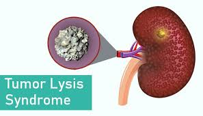
Introduction:
Tumor lysis syndrome is a complication in which cancerous tumors break down. This complication is caused by chemotherapy and radiation therapy in which cancerous tumors break down and due to which the fluid of the cancerous tumor is released and accumulates in the body and the kidneys do not function properly and due to this, intracellular fluid collects in the body and An oedematous condition occurs. And this condition is very fatal i.e. harmful.
Etiology:
- Immidiatly cell breakdown after chemotherapy
- High-grade lymphoma (such as Burkitt lymphoma)
- Acute leukemia (ALL, AML)
- Large or rapidly growing tumor
- After radiation therapy
- CAR-T or Targeted Therapy
- Ureter disease or dehydration
- Spontaneous TLS – very rare, tumor cells break down without treatment
clinical manifestation (symptoms and signs):
- fatigue,
- weakness( weakness ),
- memory loss ( memory loss ),
- altered mental status( alter mental status ),
- muscles cramp ( muscle cramps ),
- tetanus( tetanus ),
- seizure( seizure ),
- Elevated blood pressure( elevated blood pressure ),
- dusrrhythmias( dysrhythmia ),
- cardiac arrest,
- anorexia,
- nausea,
- vomiting,
- abdominal cramps,
- diarrhea( diarrhea),
- oliguria( oligouria),
- anuria( anuria),
- Renal failure( renal failure),
- acidic urine pH( acidic urine pH).
Diagnostic evaluation (Diagnostic Evaluation):
- History tacking and physical examination (History Taking and Physical Examination),
- laboratory investigation(Laboratory Investigations).
Management (Management):
- Provide diuretic medicine to the patient.
- Administer hypertonic dextrose and regular insulin. It moves potassium into cells and reduces potassium levels.
- Identify patients who are at high risk.
- Maintain the patient’s hydration status.
- Check the patient for any electrolyte imbalance.
- Check the patient’s urine pH Assess.
- Monitor the patient’s fluid and electrolyte balance.
- Maintain the patient’s hydration status.
- Provide psychological support to the patient.

FOR UNLOCK 🔓 FULL COURSE NOW. MORE DETAILS CALL US OR WATSAPP ON- 8485976407.
To unlock the complete course 🔓 or for more information, contact or whatsapp the following number.-
8485976407.