ENGLISH ANATOMY UNIT 6 RESPIRATORY SYSTEM
INTRODUCTION
For the human body to be alive, every cell of the body needs nutrient material and oxygen to perform its normal functions. Human body cannot survive without it.
Respiration and its types.
Respiration means gas exchange between any two surfaces. In respiration, oxygen enters the body from the atmosphere with inspiration and carbon dioxide accumulated in the body as waste leaves through expiration. Thus the action of respiration is observed.
Two main types of respiration are found in the body.
The exchange of oxygen and carbon dioxide between each cell tissue of the body and the surrounding blood capillaries is called internal respiration. This respiration is found everywhere in the whole body.
The gas exchange of oxygen and carbon dioxide between the lung tissue i.e. alveoli and the surrounding blood capillaries is called external respiration. This respiration is seen in the lungs.
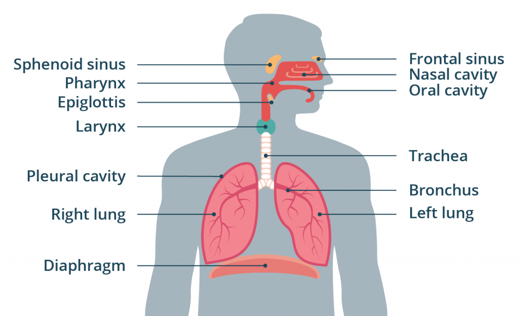
List out the organs of the respiratory system.
Following is the list of organs of respiratory system.
Organs located above the respiratory tract outside the thoracic cavity are called organs of the upper respiratory tract. Which includes the following organs.
Nose
Pharynx
Larynx
The organs of the respiratory tract located within the thoracic cavity are called the organs of the lower respiratory tract. The organs are as follows.
Trachea
Bronchi (Right and Left)
Bronchioles
Terminal Bronchioles
Alveoli
Lungs (Right and left).
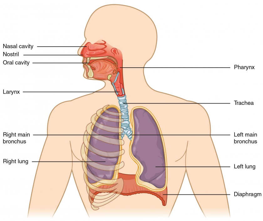
Structure of Nose and its function.
The nose is an organ at the beginning of the respiratory system. Which is located at the front of the face.
Two openings are seen from the outside of the nose. Which is called external nurse or nostril. Between these two openings is a partition, anteriorly the septum made of cartilage and posteriorly the vomer bone is the structure that separates the two openings.
The external nose is covered with skin on the outside and the inner lining is made of mucus membrane.
It contains pseudo stratified ciliated epithelium cells. There are goblet cells, which secrete mucus. Due to which the inner lining remains moist.
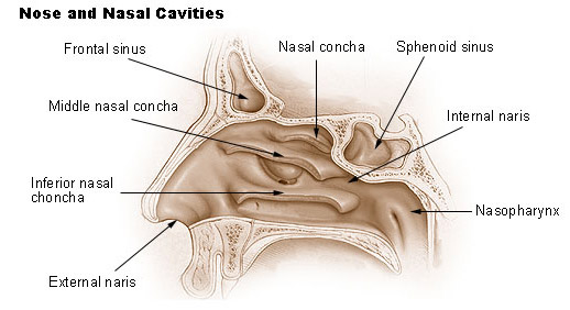
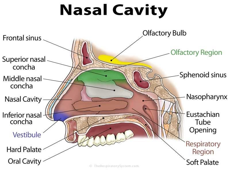
The nasal cavity is a cavity on the face, arranged above the oral cavity, and above the nasal cavity lies the cribriform plate of the ethmoid bone of the cranium. Apart from this, frontal bone and sphenoid bone are also found in this part.
The medial wall of the nasal cavity is formed by cartilage and vomer bone. This wall is known as the nasal septum.
On the floor of the nose are soft and hard pallets.
Nasal bones are located in the lateral wall of the nose. In addition, a bony structure called the inferior conkai also forms the lateral wall.
The openings at the back of the nasal cavity called the posterior ners are connected to the fairings.
There are hairs on the inside and anterior side of the nasal cavity, which warms the air, filters it and prevents small foreign particles from entering.
Openings into the nasal cavity.
There are 2 openings at the front of the nasal cavity called the anterior nares. 2 such openings are located on the posterior side of the nasal cvt, which opens towards the fairings. It is called posterior nurse.
In addition to this, the sinus opening of the bone adjacent to the nasal cavity opens. It is called para nasal sinus.
- Functions of the Nose.
Nose is the external organ of the respiratory system. Its functions are as follows.
It causes respiration. In which the oxygen wadi of external environment enters the lungs through the nose and the air with the carbon dioxide waste of the body exits through the nose.
Hairs in the inner lining of the nose clean the air. So that no foreign particles enter the respiratory tract.
Due to the hair and vascular mucus membrane in the lining of the nasal cavity, the incoming air becomes warm and reaches the lungs as warm as the body temperature so that the lung tissue is not irritated or damaged.
The inner membrane of the nose is a moist membrane due to the presence of goblet epithelium cells. As air passes through it, it becomes moist. So that the lining of the inner mucus membrane is not damaged or irritated. Thus it also performs the function of humidification.
A vestibule structure at the beginning of the nose which is upturned and has hairy processes. It works to filter the incoming air. Prevents dust particles and foreign materials from entering the respiratory tract.
The nose senses the smell i.e. it also performs the function of smelling. The receptors of the olfactory nerve are located in the mucus membrane of the nose. When the air enters the nasal cavity, the receptors are stimulated by exposure to odorous or odorous chemicals, and its impulses travel through the olfactory nerve to the brain and the sensation of smell occurs. Thus it also acts as a sense of smell.
- Paranasal sinuses and its functions.
A sinus is a small foramen in the bone. There are holes in the bone surrounding the nasal cavity, which open into the nasal cavity. All these sinuses are called paranasal sinuses.
These sinuses are air-filled cavities surrounded by a mucus membrane
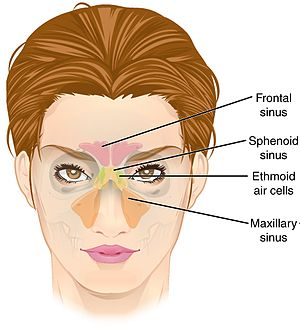
Maxillary sinuses.
Frontal sinuses.
Ethmoidal sinuses.
Sphenoid sinuses.
All the above bones have two sinuses. Each of these sinuses opens into the major cavity.
Functions of Sinuses.
These sinuses create an air field cavity in the bone due to which the bone becomes lighter in weight.
Makes the skull and face lighter.
Moistens and humidifies the incoming air during inspiration.
Maintains nasal cavity air pressure.
Recognizes surrounding sounds.
Also useful for the sensation of smell.
Structure and function of Pharynx.
Fairings are a tube like structure. Which is located in the upper respiratory tract. It is 12 cm long and 3.5 cm wide.
It is arranged behind the nose, oral cavity and larynx. It lies from the base of the occipital bone to the level of the 6th cervical vertebra.
Fairings are divided into following parts based on the structure around it.
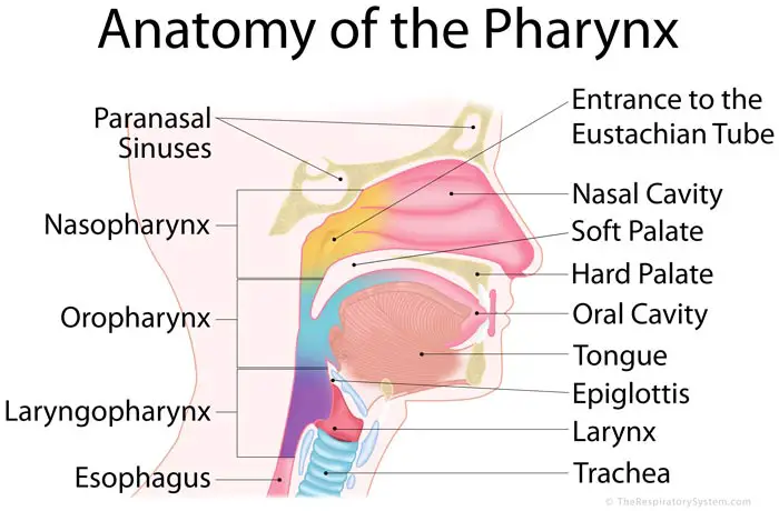
Nasopharynx.
Nose fairings means the part of the rear fairings of the nasal cavity. The 2 openings of the posterior nares behind the nasal cavity open to the nasal fairings. Another opening also opens here, in which the 2 openings of the Eustachian tube from either side of the middle ear also open here. Apart from this, 1 opening of oropharyngeals is also seen in this part. So, total 5 openings are seen on the side of Nezo fairings.
Due to the connection of this part with the middle ear, air pressure is maintained between the fairings and the middle ear.
The wall of nose fairings is rigid and thick. Hence it does not shrink. So it helps to maintain a patent airway.
There is a mass of lymphoid tissue on the upper wall of the nasal pharynges. Which is called pharyngeal tonsil.
Ciliated columnar epithelium tissue is found lining this part.
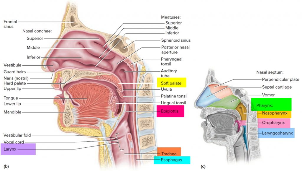
Oropharynx.
The fairings at the back of the oral cavity are called oro fairings. It is attached to the nose fairings at the top and forms the back of the mouth. Below it begins the Larigo fairings.
Part of the aurora fairings is seen from the level of the soft palate to the level of the 3rd cervical vertebra, where the hyoid bone rests.
Auro pharyngs have masses of lymphoid tissue on both sides of its wall. It is called palatine tonsil. This helps in providing local protection.
A portion of the aurophytes is composed of non-keratinized stratified squamous epithelium tissue.
Laryngopharynx.
The parts of the fairings adjacent to the laryngs are known as laryngo fairings. It is the part that starts at the level of hyoid bone i.e. from 3rd to 6th cervical vertebra.
On the upper side it is connected with the oro-farings and on its lower side the opening of the esophagus is seen.
Both the oro pharynges and the laryngo pharynges act as pathways for both the respiratory and digestive tracts.
Structural Layers of Pharynx.
The fairings are a structure made up of muscle and tissue. Which is connected with the function of both respiratory and digestive system.
The structure of the fairings consists of three tissue layers arranged from outer to inner. Which are as follows.
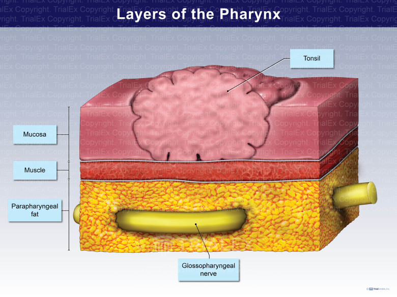
Muscular tissue layer.
It is a tissue layer made up of muscles on the outermost side of the fairings. It is a layer made up of involuntary as well as some voluntary muscles. Most of the muscles are of the involuntary type. This helps in the muscle loosening process.
Fibrous tissue layer.
This is the middle layer in the fairings. Which is a layer made up of many collagen fibers and connective tissue. This layer is thickest near the nasal pharynges and decreases in thickness towards the oropharynges and the laryngopharynges. Hence the arch portion of nose fairings remains patent airway, does not collapse.
Mucous membrane layer.
The innermost lining of the fairings is made up of a layer of mucus membrane. Ciliated columnar epithelium tissue is found in naso pharyngs and stratified epithelium tissue in oro pharyngs and laryngo pharynges.
This layer forms the continuous inner lining of the mouth, pharyns, larynx, and esophagus.
Functions of Pharynx.
It helps in the hearing process by balancing the air pressure between the fairings and the middle ear as it is attached to the Eustachian tube near the nose fairings.
Helps to warm the air taken in inspiration. Also makes it humidifying. So the air entering the lungs becomes moist and warms up to the body temperature. So the tissue of the inner lining is not damaged.
Due to the presence of lymphatic tissue cells i.e. tonsils near the pharyngs, it helps in providing specific defense against micro organisms. Thus it also performs the function of protection.
Since the endings of the olfactory nerve are located near the fairings, it is also important to understand the test.
Due to the elasticity of the fairings it also helps in producing sound quality. It works for speech too.
The pharyns act as pathways for both the respiratory and digestive tracts. So it is important in both air passage and food passage function.
Structure and function of larynx.
The larynges are structures made of cartilage. which is arranged at the bottom of the fairings and connected to the Trakia below it.
The larynx is also known as the second voice box, as it is the organ that produces sound.
It is located from the side of the hyoid bone i.e. at the level of 3rd cervical vertebra to 6th cervical vertebra.
The structure of the larynges mainly consists of cartilage. This cartilage is made up of chondrocyte cells.
The following cartilages in the structure of laryngs
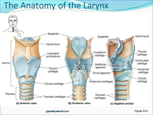
Unpaired cartilages.
There are some cartilages that have a single structure in the structure of laryngs, which are as follows.
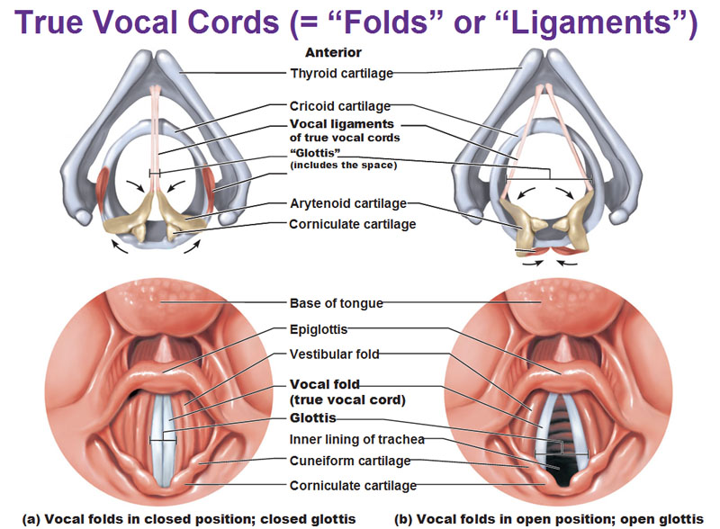
Thyroid Cartilage.
The largest cartilage in the structure of the larynx is the thyroid cartilage, which forms the V-shaped structure of the larynx. This part is made of hyaline cartilage.
In this cartilage, the subcellular cartilage portion of the thyroid on both sides is known as the thyroid lamina and the grooved portion in between is known as the thyroid notch.
The processes extending superiorly and inferiorly from the thyroid cartilage lamina are known as superior and inferior cornu of thyroid cartilage.
This raised portion of thyroid cartilage is also known as Adam’s apple.
Cricoid cartilage.
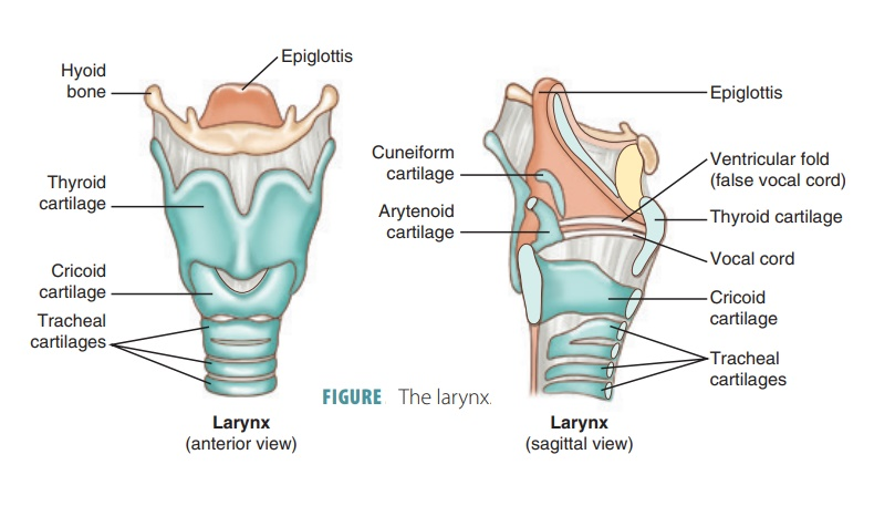
This cartilage is arranged like a signet ring on the underside of the thyroid cartilage. Its shape is seen as a truncated ring shape. This cartilage is also made of hyaline cartilage.
The cricoid cartilage is connected to the trachea and thyroid cartilage by a ligament.
When a tracheostomy procedure is performed, the cricoid cartilage is considered as a landmark.
Epiglottis cartilage.
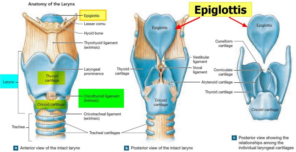
This is a leaf shaped structure. Which is made of cartilage. Which is located on the upper side of the thyroid cartilage. The upper part is wide and the lower part is narrow.
During the dissolving process it moves to the larynx and esophagus. This prevents food and liquids from entering the larynges and trachea. Elastic cartilage is present in its structure.
Paired cartilages.
In the structure of layers, some cartilages are in pairs. These types of cartilage are as follows.
Arytenoid cartilage.
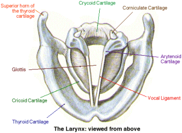
It is made of hyaline cartilage. It forms part of a triangular shape. which is attached to the vocal cords and its base is attached to the cricoid cartilage.
Corniculate cartilage.
It is a horn-shaped cartilage made of elastic tissue. It is attached to the anterior part of the arytenoid cartilage. It is the smallest cartilage among all cartilages.
Cuneiform cartilage.
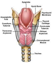
It is a rod-shaped cartilage of elastic tissue near the base of the epiglottis. It is the supporting structure of the epiglottis.
It lies anterior to the corniculate cartilage and supports the vocal cords.
Blood supply to the larynx is through the laryngeal artery and venous return is through the laryngeal vessels. The nerve is supplied by the laryngeal nerve.
Production of voice.
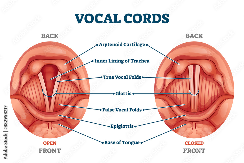
Within the laryngs, the layer of mucus membrane forms a pair of two folds. In which the superior pair is called false vocal cord or ventricular fold and the inferior pair is called true vocal cord or vocal fold.
The space between the two ventricular folds is called the rima vestibuli and the space between the two vocal folds is called the rima glottidis.
Both vocal folds straighten and vibrate when air pressure is released from the thoracic cavity. Due to which sound is produced. High pitch sound is produced due to applying more pressure and tension. Whereas low pitch sound can be produced due to less tension and pressure.
The quality of sound depends on the structure of oral cavity, teeth, nasal cavity, pharyngs, sinuses, vibrating capacity of vocal cord etc.
Functions of Larynx.
The main function of the larynx is to produce sound.
The characteristics of sound include pitch, resonance, volume etc. All these things are based on the structure of oral cavity, pharyngs, larynx, nasal cavity, sinuses, tongue and teeth as well as lips etc.
Larynx acts as an air passage.
The inner lining of the larynges contains mucus membrane and cilia which act to filter the air, warm the air to body temperature and also act in humidifying the air. Due to this, damage to the inner mucus membrane stops.
All of the above organs are considered organs of the upper respiratory tract, as they are structures outside the thoracic cavity.
Organs within the thoracic cavity are considered organs of the lower respiratory tract. The organs involved are as follows.
Structure and Function of Trachea.
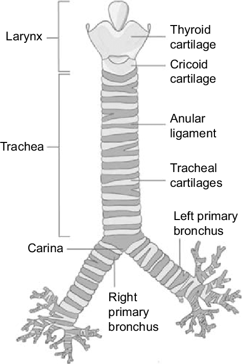
Trachea is a pipe like structure. The larynges start from the posterior part and extend up to the bronchi.
It is also known as wind pipe. This acts as the principal air passage. Its length is 12 cm and its diameter is found to be 2.5 cm.
The lining of the trachea is made of C-shaped ring-like cartilage. These cartilages are located in the anterior wall of the trachea. There are no such cartilages in the posterior wall.
The anterior wall of the trachea is composed of 16 to 20 incomplete C-shaped cartilages of this type. This cartilage increases the strength of the anterior wall. So that the trachea does not collapse due to pressure from the front side and this cartilage acts to protect it from the front side. These cartilages are of hyaline type.
Surrounding the trachea are 4 tissue layers arranged as follows.
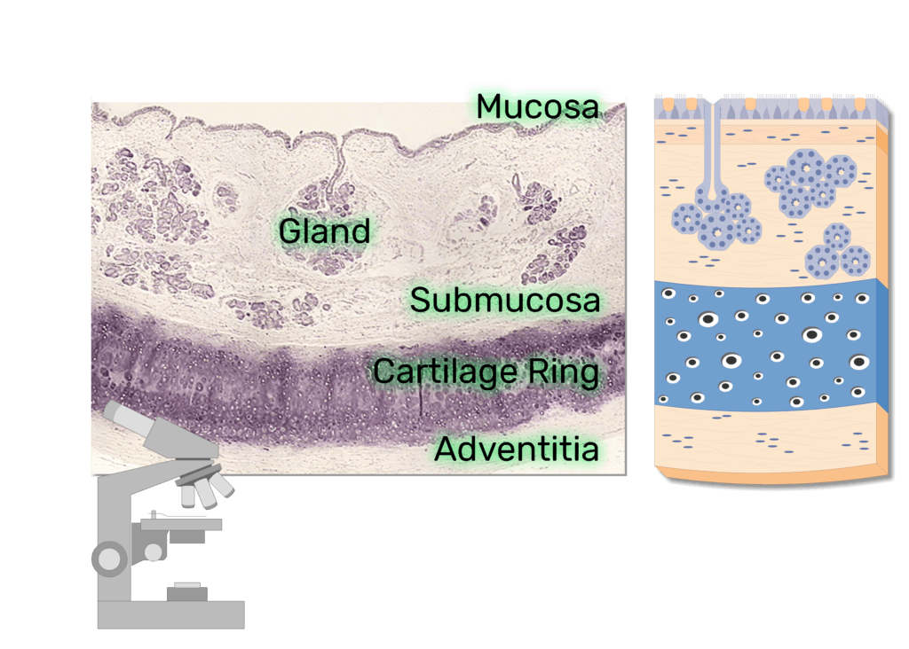
Adventitia
This tissue layer is located on the outermost side of the trachea. It is composed of areolar connective tissue.
Hyaline cartilage.
On the underside of the adventitia layer are these hyaline cartilages. Fibroelastic tissue is present in this layer and C-shaped hyaline cartilage is arranged in the anterior wall of the trachea.
Submucosa.
This layer lies beneath the hyaline cartilage. Which is the layer arranged on top of the mucosa layer. Mucus glands and their ducts are located in this layer. Apart from this, blood vessels, lymph vessels and nerves are arranged in this layer.
Mucosa.
This layer is the innermost wall of Thrace. which is composed of ciliated columnar epithelium tissue. Goblet cells are also present in this layer. which secretes mucus. This layer is moist.
This layer helps in humidifying, filtering and warming the air entering the lungs.
Functions of Trachea.
It acts as the principal air passage.
It helps in warming, filtering and humidifying the air entering the lungs.
With the help of the cartilage in the front wall of the trachea, the opening inside the trachea remains open, it does not collapse and the patency of the airway is maintained.
Cilia in the inner lining of the trachea are arranged in the reverse direction and help to move the contents, mucus or dust particles etc. deposited inside the trachea to the outside. So that the airway can be kept clear.
Nerve endings are located in the inner wall of the trachea. Which is sensitive to any irritation. Any irritation to the inner lining of the trachea stimulates the respiratory center. So the cough reflex is produced and mucus and foreign material deposited inside can be expelled by cuffing.
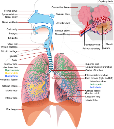
Structure of Bronchi and Bronchioles.
At the level of the 5th thoracic vertebra, the trachea divides into two branches. The branch is called bronchi. Right and left bronchi which are called primary bronchi. The branches of the bronchial tree begin from here
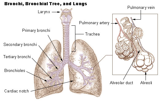
Right Bronchi.
The right bronchi are shorter and wider than the left bronchi and are arranged in a more vertical shape. So there are chances of foreign body getting more plugged in the right bronchi.
It is found to be approximately one inch long. Ciliated columnar epithelium tissue is arranged in its wall. Its wall is made of incomplete cartilage.
The right bronchus further divides into three different branches in the three lobes of the lung and one branch enters each lobe of the lung. Then it enters the lobes of the lungs and divides into small branches. It is divided into secondary and tertiary bronchi and bronchioles.
Tertiary bronchi are transformed anteriorly into grapevine-like alveoli. There are millions of alveoli in the structure of the lungs. As we move towards the alveoli in the bronchial tree, the cartilage disappears.
Left Bronchi.
The left bronchus is narrower and longer than the right bronchus. Its length is approximately 2 inches. Its structure is also similar to right bronchi. It forms 2 branches and enters the two lobes of the left lung going forward and further divides into many branches in each lobe of the lung.
Oxygenated blood is supplied by the bronchial arteries and deoxygenated blood is returned by the bronchial veins. It is supplied with nerves by the autonomic nervous system.
Functions of Bronchi and Bronchioles.
It works by warming and humidifying the air.
It stimulates the cough reflex so that the respiratory tract is protected.
It keeps the track patent so that respiration can be taken with ease.
It works to remove the mucus accumulated in the bronchi through the cough reflex.
The entry control of the air taken in breathing makes equal distribution everywhere.
Structure of Alveoli.
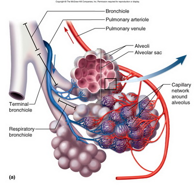
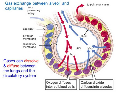
At the ends of the terminal bronchioles are tiny air sacs like grape skins. Which is called alveoli. This is the primary structure for gas exchange to occur in the lungs.
The wall of alveoli is very thin and composed of single squamous epithelium tissue. So it can be easily done by gas exchange diffusion method of oxygen and carbon dioxide.
Surrounding these alveoli is a network of pulmonary blood capillaries. This gas exchange between blood and alveoli is also called external respiration.
Fluid is present on the surface of the alveoli. Which is called surfactant. This surfactant reduces the surface tension. Prevents alveoli from collapsing. Prevents alveoli membrane from drying and also helps in gas exchange.
Functions of Alveoli.
The main function of the alveoli is gas exchange. It carries out gas exchange of oxygen and carbon dioxide.
It humidifies and warms the air as it surrounds the pulmonary capillaries. Due to this, the air becomes warm and moist.
As the membrane of alveoli is wet, it gets plugged with dust particles and foreign materials and also works to remove them through cuffing and sneezing.
Connective tissue is present in the wall of alveoli. So it also contains lymphocytes and plasma cells and it also synthesizes some antibodies, which provide resistance to local foreign bodies and act as protection.
- Structure of the Lung.
Lung is an important organ of respiratory system. They are located on either side of the mediastinum space in the thoracic cavity, totaling 2 in number.
The lungs take in oxygen from the atmosphere and expel carbon dioxide from the body.
Lungs are located in the thoracic cavity in number of 2. They are conical in shape.
The lungs are separated from the heart and the thoracic cavity by the mediastinum space.
The lungs are made up of spongy tissue within which many air field cavities are located. Its color is brown or grey.
The weight of the right lung is approximately 625 grams and the weight of the left lung is approximately 575 grams. The right lung is heavier in weight and larger in structure than the left lung.
Lungs are divided into lobes. The right lung has 3 lobes, namely the superior lobe, middle lobe and inferior lobe, while the left lung has 2 lobes, the superior lobe and the inferior lobe. These lobes are separated by a fissure. There are two fissures in the right lung. A fissure is seen in the left lung.
Lungs are classified into the following parts.
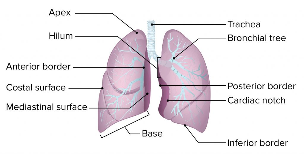
. Apex..
The upper triangular and round part of lunge is called Apex.Which is seen up to the level of the clavicle bone.
- Bayes.
The lower broad part of the lung is called the base. The base portion is attached to the diaphragm at the bottom. This part is of concave shape.
- Anterior border..
It is thin. It is shorter than the posterior border. It has a cardiac notch. In which the heart part is arranged.
- Posterior border..
It is thick. It is found from the 7th cervical vertebra to the 10th thoracic vertebra.
- Inferior border..
It is located at the bottom of the lung. It separates the costal surface and the medial surface. The costal surface is large and convex. It is in contact with the costal pleura. It is attached to the ribs and intercostal muscles by costal cartilage.
- Medial surface
It is concave. There is a groove in the middle of it which is called the hilum. The hilum lies at the level of the fifth, sixth and seventh thoracic vertebrae. Through this hilum, bronchi, pulmonary blood vessels, lymphatic vessels and nerves enter and exit each lobe of the lung.
In the middle of the medial surface lies the mediastinum space. which separates the two lungs. In this space there are structures like heart, great vessels, trachea, bronchi, esophagus etc. which separate both the lungs.
Structure of the Lobe of the Lung..
The lobes of the lungs are made up of many lobules. One lobe is separated from the other lobe by a fissure. In the center of the lung is a groove called the hilum. From this hilum the following structures are found in each lobe.
Bronchi enter from each lobe of the lung. After entering, it divides and transforms into secondary bronchus, tertiary bronchus, terminal bronchioles, alveolar shakes and small grape-like alveoli. Thus, this structure is seen in a tree-like structure in the lobes of the lung, which is called bronchial tree or respiratory tree.
Surrounding these alveoli is a network of capillaries of the pulmonary artery and pulmonary vein. Gas exchange takes place here between oxygen in the alveoli and carbon dioxide in the blood capillaries through inspiration. This is known as external respiration.
Thus, in each lobe of the lung there is a network of bronchial tree, capillaries of pulmonary vessels, lymph capillaries, nerves and parenchymal tissue of the lung.
pleura..
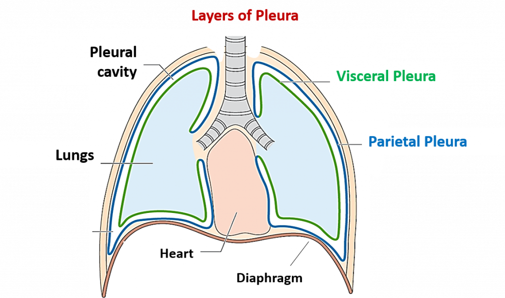
The pleura is the serous membrane surrounding both lungs. Which is found in double layer. The outer layer is known as parietal pleura and the inner layer is known as visceral pleura.
Between the parietal pleura and the visceral pleura lies a cavity called the pleural cavity. There is serous fluid which is also called pleural fluid.
Due to the pleural fluid in the pleural cavity, the two layers do not rub against each other and due to this, the lungs get enough space for expansion. The pleural fluid in this cavity is viscous and also acts as a lubricant.
The visceral pleura is the layer adjacent to and adjacent to the lungs.Whereas the parietal pleura is the layer attached to the ribs and muscles.
Difference between Right Lung and Left Lung..
Right lung has two fissures to separate the lobes while left lung has only one fissure to separate the lobes.
Right lung has three lobes superior, middle and inferior while left lung has two lobes superior and inferior.
The right lung has a straight anterior border while the left lung has an interrupted anterior border because of the cardiac notch on the side of the left lung where the heart is located.
Right lung is heavier and larger in weight. Its weight is approximately 625 grams. While the left lung is lighter and smaller in weight. Its approximate weight is 575 grams.
The right lung is short and wide while the left lung is long and narrow.
Function of Lung..
It is an important organ for respiration.
Through inspiration, oxygen reaches the lungs and mixes with blood to deliver oxygen to the whole body.
The waste product of the body, carbon dioxide, is removed from the body through the act of exhalation.
Oxygenated blood is delivered to the heart through the pulmonary circulation.
The body excretes excess water through the act of respiration.
Other gaseous wastes produced in the body are removed from the body through the act of exhalation.
- Respiratory muscles.
Respiration means bringing the outside air to the lungs through the act of inspiration and throwing the air accumulated inside the lungs to the outside atmosphere through the act of exhalation.
During this action, there is an increase and decrease in the size of the thoracic cavity, which is mainly done by the muscles associated with the act of respiration.
The muscles of respiration perform mainly involuntary work. The muscles involved in this action are as follows.
Diaphragm.
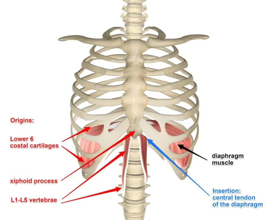
Located between the thoracic cvt and the abdominal cvt, this muscle has a dome shape structure.
The diaphragm receives contraction impulses via the phrenic nerve. So its movement up and down is seen.
The diaphragm is attached to the thoracic vertebrae by a central tendon. It is also attached superiorly to the scyphoid process, pleura and side ribs. Hence, due to the movement of the diaphragm, there is a fluctuation in the movement of the chest cavity.
During the act of inspiration, the diaphragm moves downwards and the increase in the size of the thoracic cavity shows the act of inspiration, when the diaphragm goes back to its original position and relaxes, the diameter of the thoracic cvt decreases and the act of exhalation is seen.
Intercostal muscles.
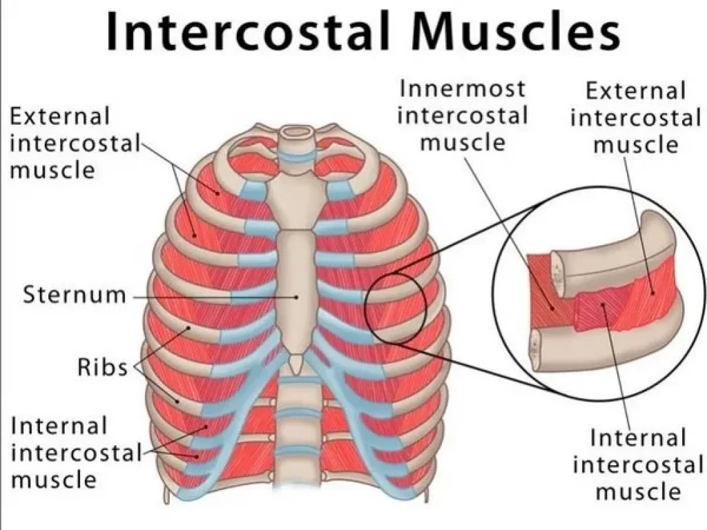
The space between two ribs is called intercostal space. The muscles attached to this part are called intercostal muscles. A total of 11 pairs of intercostal muscles are found. It is arranged in two layers. On the outer side are the external intercostal muscles and on the inner side are the internal intercostal muscles.
Impulses are supplied to these intercostal muscles by the intercostal nerve. The first rib is fixed and the rest of the ribs move towards the first rib due to the movement of the intercostal muscles.
When the intercostal muscles contract, they increase the diameter of the CVT and when they relax, the thoracic CVT decreases in size.
The action of inspiration and expiration occurs from the contraction and relaxation of both the above muscles diaphragm and intercostal muscles.
- Cycle of respiration (Mechanism of respiration).
Respiration is gas exchange between two surfaces. In which air from the atmosphere enters the lungs. Gas exchange between lung tissue and blood is called external respiration. The gas exchange that takes place between each cell tissue of the body and the blood is called internal respiration.
During the act of respiration, oxygen enters the lungs through inhalation and carbon dioxide leaves the body through exhalation.
Normally, the act of respiration is observed 16 to 18 times in a minute.
The following steps are observed in the cycle of resuspension.
Inspiration
expiration
pos.
Inspiration (Inspiration).
Inhalation of atmospheric air into the lungs is called inspiration.
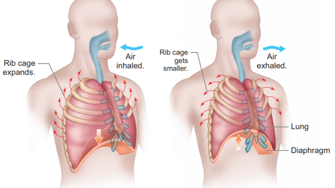
When the brain receives nerve impulses for contraction of the diaphragm and intercostal muscles, the contraction of the diaphragm and intercostal muscles increases the size of the thoracic cavity. The air pressure inside the thoracic cavity decreases so that air from the outside atmosphere can enter the lungs through the action of inspiration. This action is called inspiration.
Contraction impulses to the diaphragm cause the diaphragm to flatten downwards and contraction of the intercostal muscles causes the ribs and intercostal muscles to move upwards and outwards. So the size of the thoracic cavity increases and negative pressure is created inside the cavity. As the air pressure in the outside environment is higher and the air pressure in the thoracic cavity is lower, the act of inspiration can occur. The act of inspiration is an active process.
Expiration (Expiration).
The process of expelling air from the lungs into the atmosphere is called exhalation. The action of expiration is a passive process that begins after the action of inspiration is completed.
The act of exhalation relaxes the contracted diaphragm and intercostal muscles. So the diaphragm returns to its original position and the ribs come down and inwards, reducing the size of the thoracic cavity and exhalation takes place. In which the air from the lungs is thrown out into the atmosphere.
In the act of exhalation, the air pressure in the lungs is greater than the atmospheric pressure so that the act of exhalation takes place.
Pause.
This is the relaxed stage of the lung. In which no action of inspiration or expiration takes place. This period is called pause period.
- Terminologies related to lung volume and vital capacities of the lung.
The action of inspiration and expiration is called respiration. Normally 12 to 18 respirations are seen in 1 minute. A spirometer or respirometer is a useful device for measuring air volume during respiration. The terminologies related to this normal respiration and lung capacity are as follows.
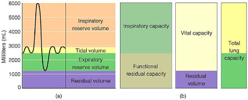
Tidal volume.
The volume of air that moves into the lungs during normal inspiration and the volume of air that moves out of the lungs during normal expiration is called tidal volume during one respiration. Normally it is around 500 ml.
Inspiratory reserve volume. (IRV).
The volume of extra air that can be inspired in addition to the air taken in during normal inspiration in the lungs is called the inspiratory reserve volume. Its normal volume is 3100ml.
Total inspiratory capacity.
Total inspiratory capacity in lung i.e. the total of Titral Volume and Inspiratory Reserve Volume is called Total Inspiratory Capacity. 500 + 3100 = 3600 This is the total inspiratory capacity of the lungs.
Expiratory reserve volume. (ERV)
The maximum volume of air that can be exhaled during forced expiration in addition to the air exhaled during normal expiration is called expiratory reserve volume. It is approximately 1200ml.
Residual volume.
The volume of excess air still remaining in the alveoli during expiratory reserve volume, which is not expelled even during forced expiration, is called residual volume.
That volume of air cannot be measured even with a spirometer and this residual volume does not cause alveoli to collapse. Its normal volume is 1200ml.
Vital capacity of lung.
The sum total of tidal volume, inspiratory reserve volume and expiratory reserve volume is called the vital capacity of the lung. It is approximately 4800 ml.
Anatomical dead space.
The volume of air in the lower respiratory tract that is not involved in gas exchange. All that space is called anatomical dead space. The air in this space acts only by conduction. Gas exchange does not work.
Total lung capacity.
This includes the vital capacity of the lung as well as the residual volume.
Total lung capacity is found to be approximately 6000ml.
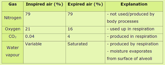
- Composition of inspired air and expired air.
Control of Respiration.

Respiration is an involuntary process. Which is not under one’s own control.
The process of rhythmic respiration is under the control of the brain. This control has the following structure.
The respiratory center in the medulla oblanta of the brain controls the rate and death of respiration.
The pneumotaxic area and the apneustic area are located near the pons veroli. Co-ordination of respiration is also maintained through it.
Chemo receptors are located in the medulla oblongata of the brain. These chemo receptors monitor the amount of carbon dioxide and oxygen in the blood there.
When there is an increase in the amount of carbon dioxide in the blood, these chemo receptors are stimulated, that is, in the condition of hypercapnia, this chemo receptor is stimulated and increases the rate of respiration.
Thus all the above structures control respiration. So the amount of oxygen and carbon dioxide in the body is maintained.