ENGLISH ANATOMY UNIT 3. THE BLOOD
- Blood..
Blood is a very important component in our body. A person’s life is not possible without blood.This is a type of connective tissue.
Blood circulates in the blood vessels in our body. With the continuous pumping of the heart, the circulation of this blood starts in the body.
Blood is a liquid that makes up 7 to 9% of our body weight. That is, in a healthy adult person, it is found as 4 to 6 liters.
Blood is a red liquid. The color of oxygenated blood is bright red due to mixing of oxygen inside the blood. When there is absence of oxygen in the blood i.e. presence of carbon dioxide, the color of blood is dull red or less red.
Blood is more dense than liquid which is called its viscosity. It ranges from 3.5 to 5.5.
The specific gravity of blood is 1.045 to 1.065.
Blood has slightly alkaline characteristics. Which has a ph of 7.35 to 7.45. Due to the lack of carbon dioxide molecules in arterial blood, it is more alkaline than venous blood.
- Composition of blood..
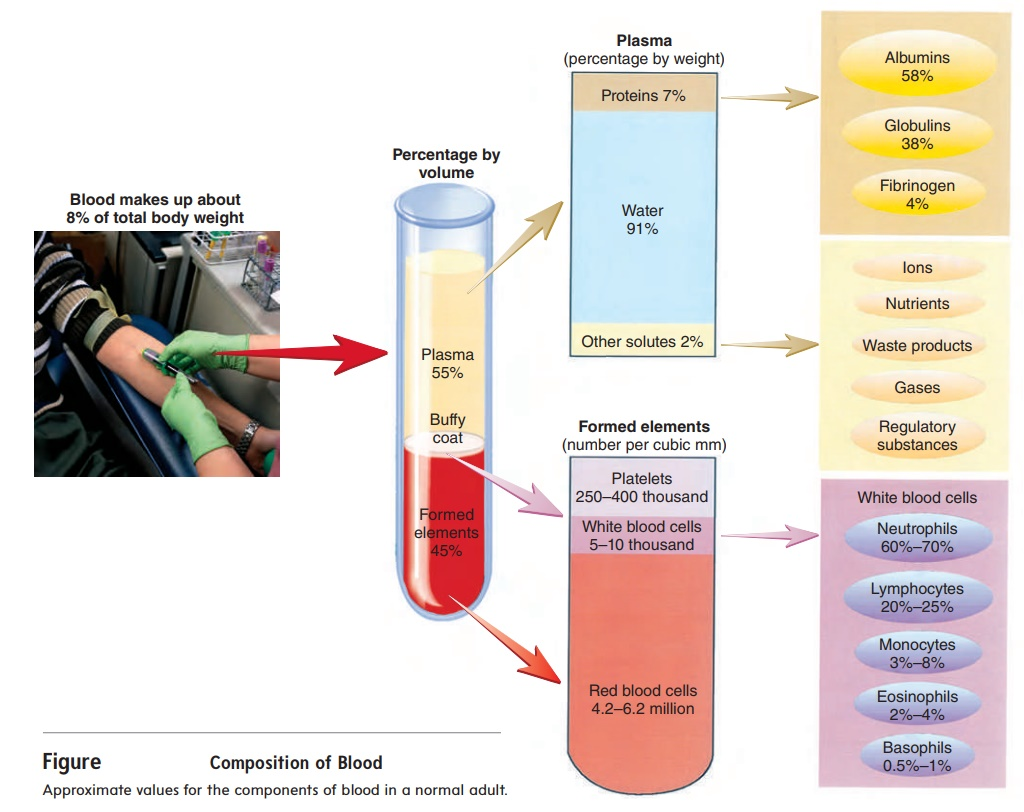
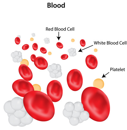
Blood is a type of connective tissue. Its structure consists of 55% plasma and 45% blood cells.
When the blood is taken out of the body and collected in a tube and kept for some time, a pale yellow liquid is seen on the surface which is called plasma. Under it, a dark red liquid is seen collected which is called cell. The components of this plasma and cell are as follows.
Components in plasma.
- Water.
Plasma contains the highest amount of water i.e. about 92% water.
Water is very essential to maintain normal hydration in the body. Water plays an important role in maintaining intracellular and extracellular balance in the body. The water in the plasma easily reaches every cell-tissue through the blood and every cell and tissue can function well.
- Protein..
Plasma contains proteins called albumin, globulin and fibrinogen. Which is found in total 7% of plasma.
Albumin..
Albumin is the most abundant protein in plasma. which is formed in the liver and performs the following functions. It works to maintain the osmotic pressure of the plasma. Albumin also acts as a carrier molecule for some other substances i.e. helps in their transport.
globulin.
Globulin is a protein present in blood plasma. It is produced by the liver and has three types. In which alpha, beta and gamma globulin proteins are present in the blood. Gamma globulin acts to maintain body immunity.
Fibrinogen..
It is a protein present in plasma. It is produced by the liver and plays an important role in the blood clotting mechanism.
- Plasma Electrolytes…
Plasma contains some electrolytes. In which positively charged electrolytes are known as cations. E.g. Sodium, potassium.
Electrolytes with a negative charge are known as anions. D. T.Chloride, Sulphate, Iodine etc.
These electrolytes act as minerals and are important in maintaining the osmotic pressure of the cell.
- Nutrients.
At the end of the digestion process in the body, the absorbed nutrient materials enter the blood and mix with the blood plasma. These nutrient materials contain macro and other micro nutrients like carbohydrates, proteins, fats etc. Which every cell of the body receives through blood plasma and the cell can perform its functions.
- Gases…
Some gases combine with the blood plasma and circulate through the blood plasma within the body. In which oxygen, carbon dioxide and nitrogen are mainly present.
- Waste product.
Any waste product produced by the cell at the end of digestion in the body. These waste products combine with the blood plasma and reach the excretory organs of the body and leave the body. This waste product contains urea, uric acid, creatinine, bilirubin etc.
- Blood cells..
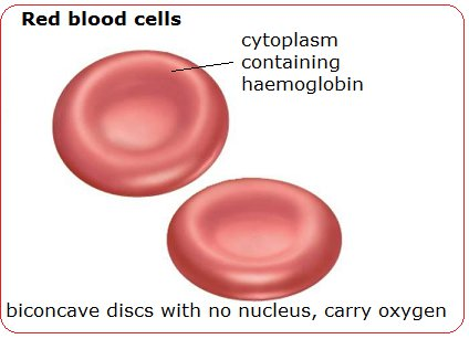
Red blood cells or erythrocytes…
RBCs make up 99% of all cells in the blood.
RBCs are circular disc-like biconcave cells without a nucleus.
Its diameter is approximately 7 microns and its thickness is 2 microns.
Immature cells in RBCs have a nucleus while mature RBCs do not have a nucleus.
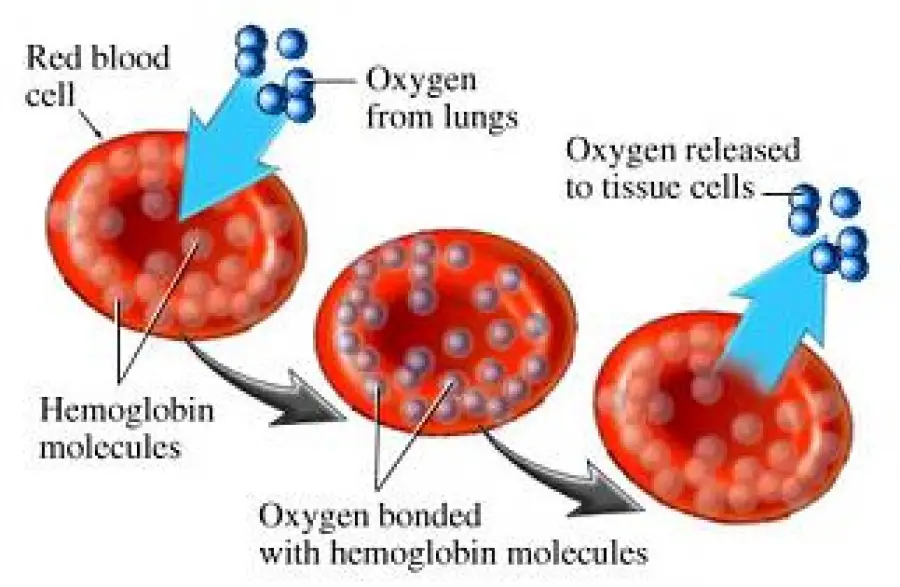
These RBCs contain a protein. Which is known as hemoglobin. Due to which its bright red color is seen. Hemoglobin and oxygen combine with RBCs to form the compound oxyhemoglobin and due to this they appear bright red in color.
Through the lungs, this oxygen enters the breath and connects with the RBCs of the blood and reaches every cell and tissue of the body. Hence the main function of RBC is to deliver oxygen to cell-tissue.
The life span of RBC is 90 to 120 days.
There are four to five million RBCs in the blood.
Production of RBC..
The process of formation of RBCs is known as erythropoiesis. It takes seven days to complete this process, and for this process, a hormone secreted by the kidneys, erythropoietin, plays a very important role.
RBCs are made from the baby’s liver and spleen during pregnancy before the baby is born. But after birth it becomes the cavity of the bone marrow of the long bone. The production of RBCs is mainly in the red bone marrow of vertebrae, ribs, sternum bone, pelvic bone, humerus and femur bone.
Certain nutrients such as amino acids, riboflavin, folic acid, vitamin B etc. are very important for the production of RBCs and their maturation. Due to the presence of these elements RBC matures.
Erythropoietin, a hormone secreted by the kidneys, is mainly responsible for the production of RBCs. The secretion of this erythropoietin hormone depends on the oxygen saturation of the blood in the artery. In which oxygen saturation decreases, this hormone is activated and this hormone stimulates the production of more RBCs. This mechanism is called negative feedback mechanism.
Destruction and Removal of RBC..

The process of distribution of RBCs is called himalayas. The life span of RBC is 90 to 120 days. Production of RBC and its distribution are maintained at the same rate.
As the RBC approaches the end of its life span, its wall becomes weaker and the process of degradation begins.
in which the RBC wall breaks and the broken RBC fragments are distributed to the body via the liver, spleen and various reticuloendothelial systems.
In which some cellular parts are cleared by the action of phagocytosis.
Globin, hemoglobin and iron are released from the RBC wall.
In which new protein synthesis takes place from the globin portion. Iron is converted to ferritin and reused during the synthesis of new RBCs.
Bilirubin and biliverdin are formed from the hemoglobin portion of RBCs. which is processed by the liver and exits the body through urine and stool.
Hemoglobin..
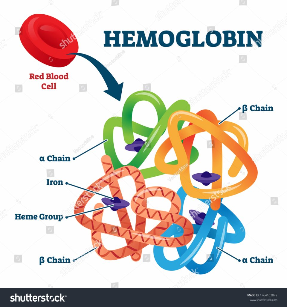
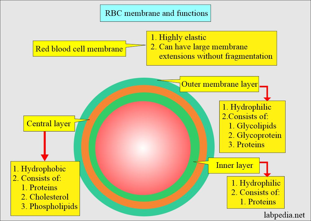
Hemoglobin is a complex protein associated with RBCs. It is located in the wall of the RBC known as the stroma. The wall of the RBC is composed of phospholipids.
In Hemoglobin, Heme i.e. Iron Portion and Globin i.e. Protein Portion combine to form Hemoglobin.
This hemoglobin contains 5% hemoglobin and 95% globin portion.
Hemoglobin in RBC binds oxygen atoms from the lungs to form oxy-hemoglobin and through RBC oxygen reaches every cell tissue of the whole body.
The bright red color of blood is due to this oxygenated hemoglobin.
The amount of hemoglobin in a person’s normal body is found to be 12 to 16 g/dl in females and 14 to 18 g/dl in males.
An increase in the number of RBCs in the blood is called erythrocytosis and a decrease in the number of RBCs in the blood is called erythrocytosis.
A condition in which the number of RBCs in the blood in the body is less than normal is also known as anemia.
Functions of RBC..
RBCs transfer oxygen from the lungs to the body and each of its cells and tissues.
Hemoglobin in RBCs carries the waste products carbon dioxide with it to the lungs and helps to remove it from the body through exhalation.
By removing carbon dioxide from the blood, RBCs also maintain the acid-base balance in the blood.
Due to the binding of oxygen to hemoglobin, oxyhemoglobin helps in giving the blood its bright red color.
- WBC..
It is the largest cell in the blood. Its main function is to provide protection to the body and destroy micro-organisms.
One percent of all blood contains WBC cells.
White blood cells are also known by other names of leukocytes. Its number in blood is 5,000 to 10,000.
An increase in white blood cell count than normal is called leukocytosis and a decrease in white blood cell count is called leukocytopenia.
This cell has a nucleus and also has the property of moving itself so that it can easily reach the site of infection. Based on the distribution of plasma granules of wbc it is divided into two types.
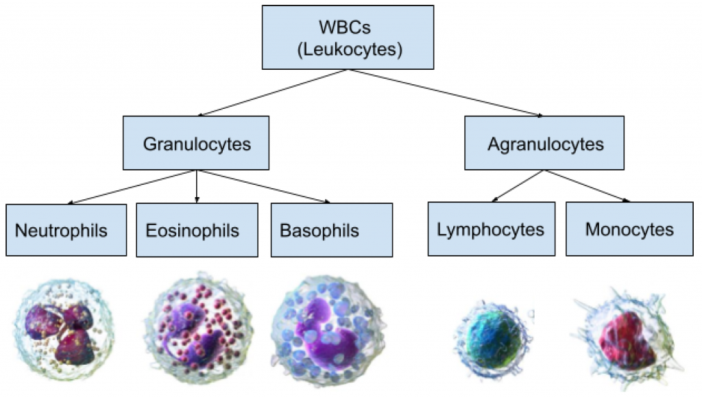
- Granulocyte.
White blood cells contain a large number of granulocyte cells. This cell has granule-like structures in its cytoplasm. It also has a multi lobed nucleus. Granulocyte White blood cells are again divided into three types.
A. Neutrophils.
It is found in 60 to 70% of total white blood cells.
It functions as a phagocytic cell. Which has the property to engulf the micro organism and destroy it in its cytoplasm.
The nucleus in the cytoplasm of neutrophils has 2 to 6 lobes.
These neutrophils act to protect the body against foreign material or micro-organisms.
It also works to remove the waste material accumulated in the body.
B. Basophils…
These cells constitute 0.5 to 1% of total wbc. Its nucleus is bi-lobed and irregular in shape. This cell contains heparin. which acts as an anticoagulant.
Basophils provide immunity to ward off parasitic infections.
C. Eosinophil…
These cells account for 2 to 4% of total wbc. It has a bilobed nucleus of B sep in its cyto plasm. This cell has phagocytic property. Its number increases during an allergic reaction. This cell contains a protein called plasminogen. A protein that helps break down fibrin in blood clots.
- Agranulocyte…
This cell contains some non-specific lysosome-type granules in its cytoplasm. They constitute 20 to 30% of the total white blood cells. This cell is divided into two main parts.
A. Monocyte…
They constitute 3 to 8% of total white blood cells. It is the largest of all WBC cells.
It contains the nucleus in the cytoplasm. It has motility properties and performs phagocytic function.
B. Lymphocyte.
They constitute 20 to 25% of the total white blood cells. It is found in small size. It contains a nucleus. It has two main types.
- B lymphocyte..
This cell originates from the bone marrow. Which is important in producing antibody against specific antigen.
- T lymphocyte…
Lymphocytes produced from the bone marrow are processed in the thymus gland and converted into T lymphocytes. These cells are very important in maintaining cellular immunity.
The production of white blood cells takes place in the bone marrow. The process of this production is known as leukopoiesis.
White blood cell production also occurs in lymphoid tissue, spleen and tonsils.
Functions of white blood cell…
The main function of WBC is phagocytosis.
Phagocytosis..
These white blood cells in our body engulf micro-organisms or foreign materials in their cyto plasm and destroy them with their own chemicals. This action is called phagocytosis.
It also works to repair certain tissues in the body.
This cell works to maintain immunity in the body.
As heparin is released by this cell, it also acts as an anticoagulant.
- Platelets…
This cell is also known by other names of thrombocytes. These cells are small in size without nucleus.
Granules are located in the cyto plasm of this cell. Its normal amount is two and a half to four lakh milliliters of blood.
The main function of this cell is to clot the blood. With its help, the process of blood clotting is done.
A higher than normal number of these cells in the body is called thrombocytosis and a lower than normal number of cells is called thrombocytopenia.
Once this cell reaches the blood stream, it stores a chemical that is also important for repairing the damaged inner lining of blood vessels.
The life span of a platelet cell is 7 to 10 days.
Hematopoiesis..
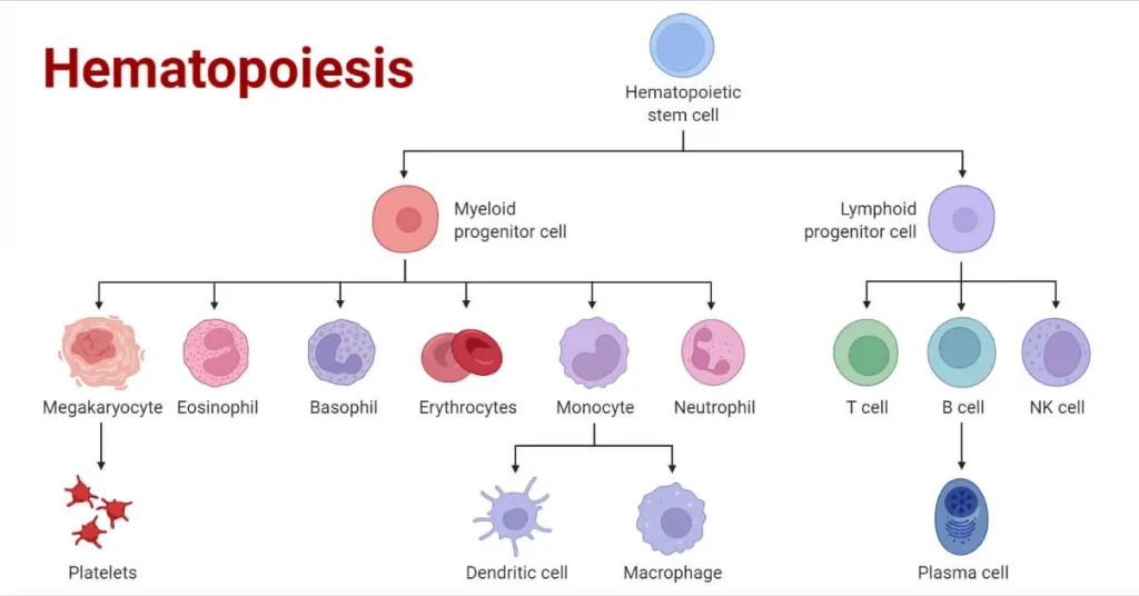
The process of formation of blood cells from bone marrow is called hematopoiesis.
Bonemarrow consists of red bonemarrow of long bone and flatbone. which is the primary site for the production of these blood cells.
Functions of Blood…
Blood is a major liquid in the body. which performs the following functions.
- Blood A blood mainly performs functions related to transportation which includes the following activities.
Blood transfers oxygen from the lungs to the body tissues and carbon dioxide from the body tissues to the lungs.
Blood transports nutrients absorbed from the alimentary canal throughout the body.
Blood carries hormones from endocrine glands to their target cells.
Transports the waste produced at the end of metabolism to the body’s excretory organs.
Helps to transport the hits generated in the body throughout the body.
- Blood acts for the regulation of certain functions in the body which are as follows.
Blood maintains the pH of both acids and bases and blood acts as a buffer solution.
Body maintains water and electrolyte balance in the blood.
Blood regulates body temperature.
- Blood also performs some protective functions which are as follows.
Due to the specific characteristics of the white blood cells present in the blood, they protect the body by providing protection against micro-organisms.
Protects the body from some toxic substances.
Due to the property of blood to clot in the cells, blood clotting mechanism prevents excess blood loss from the body.
- Hemostasis…
Haemostasis is the act of stopping blood flow. When bleeding occurs due to an injury anywhere in the body, if there is no blood clotting mechanism, then the entire body bleeds. But it doesn’t happen like that. Blood stops flowing through the action of hemostasis in the body. There are three phases in this action. Which are as follows.
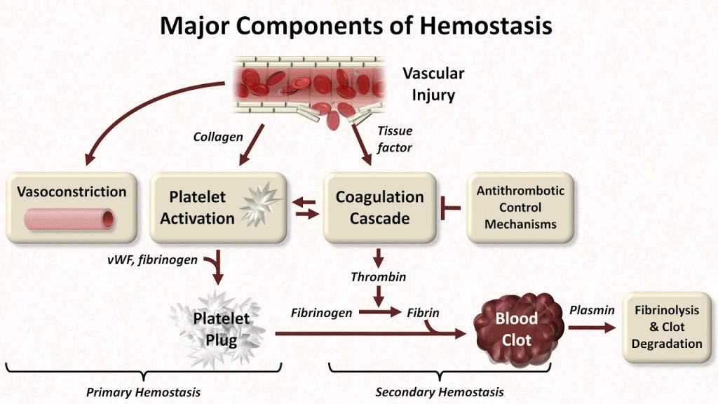
- Vaso constrictive phase…
During this phase, when blood flows out of the damaged blood vessels, chemicals are released around it and the injured smooth muscles spasm.
Muscle spasm and release of vasoconstrictive chemicals cause blood vessels to constrict and blood flow to the injured area decreases. This phase is called vasoconstrictive phase.Blood clotting is promoted during this phase due to reduced blood flow to the injured site for blood clotting.
- Platelet plug formation..
Platelets are released when blood flows from injured blood vessels. The chemical adenosine diphosphate is released from the injured site so that more platelets are attracted to it and the platelets adhere to the damaged blood vessels and act to form a temporary seal there. Due to the formation of this seal there is a decrease in blood flow.
- Blood co-coagulation or blood clotting mechanism…
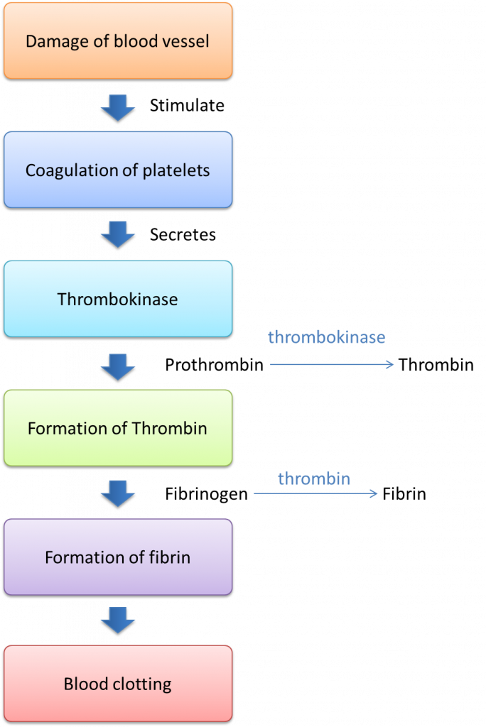
In this mechanism many chemical reactions are observed in a series. In which a chemical called thromboplastin is released from the site of damaged blood vessels. This released thromboplastin binds to calcium ions and converts prothrombin to thrombin.
This thrombin converts fibrinogen to fibrin and fibrin forms a fibrous network around the injured area. Blood cells get trapped in this network and a blood clot is formed.
Thrombin activates fibrin stabilizing factor. which gives additional strength to the fibrin strands so that the blood clot hardens and this mechanism continues until the complete stoppage of blood flow from the injured site.
- Fibrinolysis…
This is a physiological mechanism to dissolve the blood clot. In which plasminogen is converted to plasmin and that plasmin acts to break up fibrinogen strands and all the waste material of fibrin is absorbed from there by the action of phagocytosis.
The action of complete hemostasis is seen in the above four phases.
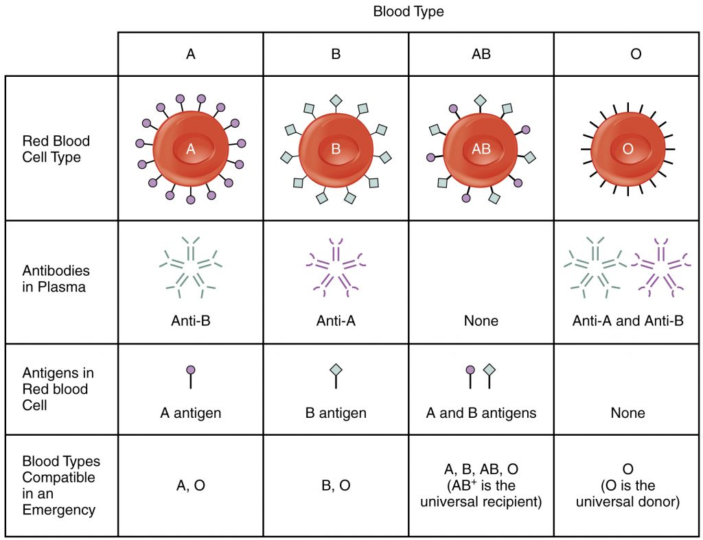
- Blood groups…
Karl Landerson discovered the blood group and divided it into four main groups.
Every person has any one of these four blood groups.
This blood group includes A, B, AB and O.
The name of this blood group is given according to the name of the antigen present in the wall of its RBC.
Antibodies other than that blood group are present in the blood plasma. If another antigen enters the body, an antibody-antigen reaction occurs and RBCs are destroyed.This action is called Himalayas.
If the donor’s blood does not match with the recipient’s blood, it is called incompatibility.
E.g. Type A blood has antigens of A and antibodies of B in the walls of its RBCs. In this way there is arrangement of antigen and antibody in blood.
AB blood group contains both A and B antigens. It does not contain any antibodies. Hence AB group is known as universal recipient.
O blood group does not contain any type of antigen A or B, it contains both types of antibodies O group is known as universal donor.
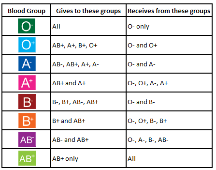
Rh System… Ris Hus System…..
Rh stands for Reisch Hus in which if this type of protein is present on the wall of RBC then it is called Rh positive person and if this type of protein is not present on the wall of RBC then it is called Rh negative person.
An estimated 80% of people are found to be Rh positive and 20% of individuals are found to be Rh negative.
If Rh positive blood is given to an Rh negative person, an immune response is created and the blood cells are destroyed i.e. himalayas. It is also called a transfusion reaction.
difference between RBC and WBC
RBCs appear red in color while WBCs are white in color or colorless.
The shape of RBC is circular biconcave disk shape.. while WBC is round and irregular shape.
Nucleus is absent in RBC.. whereas nucleus is present in WBC..
RBCs are involved in the transportation of oxygen while WBCs are involved in maintaining immunity and defense mechanisms in the body.
The life span of RBC is 90 to 120 days while the life span of WBC is few hours to years.
Its function is linked to the cardiovascular system while the function of wbc is linked to both the cardio vascular system and the lymphatic system.
RBCs constitute 40 to 45% of the total blood while WBCs constitute one percent of the total blood.
Only one type of RBC is found in blood whereas five types of WBC are found in blood.
RBCs have the property of circulating only in the blood circulation while WBCs can travel beyond the blood circulation to the connective tissue and lymphatic system when needed.
Decreased RBC than normal results in anemia while decreased in WBC results in leukopenia.